Chapter 14 The Central Nervous System
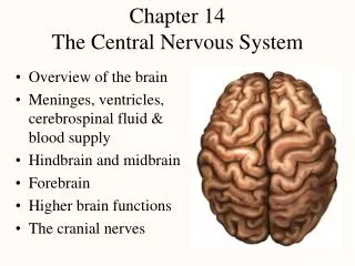
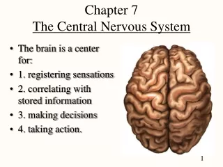
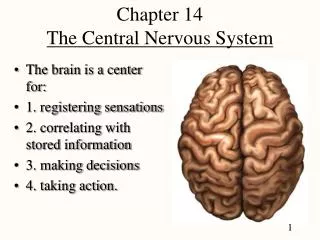
Chapter 14 The Central Nervous System. The brain is a center for: 1. registering sensations 2. correlating with stored information 3. making decisions 4. taking action. Landmarks. Major parts of the brain - cerebrum, cerebellum, brainstem

Chapter 14 The Central Nervous System
E N D
Presentation Transcript
Chapter 14The Central Nervous System • The brain is a center for: • 1. registering sensations • 2. correlating with stored information • 3. making decisions • 4. taking action.
Landmarks • Major parts of the brain - cerebrum, cerebellum, brainstem • brain weighs about 3 pounds, more in anatomy students
Lobes of the Brain • The lobes of the brain are named for the cranial bones the overlie them • The frontal lobe has the prefrontal cortex, the anterior part of the frontal lobe
Brain • Longitudinal fissure separates 2 cerebral hemispheres • Fissures are deep grooves • sulci the shallow grooves • gyri are the elevated folds • surface layer of gray matter is called the cortex, squash, noodle
Cranial Meninges • Dura mater -- outermost, tough membrane • outer periosteal layer against bone • forms dural venous sinuses draining blood from brain • supportive structure formed by dura mater • falx cerebri • Arachnoid mater is spider web filamentous layer • Pia mater is a thin vascular layer adherent to contours of brain • The brain has a PAD around it that is continuous with the _____ ____
Cerebrospinal Fluid • Clear liquid fills ventricles and canals, it flows in the subarachnoid space • Functions • buoyancy -- floats brain so it neutrally buoyant • protection -- cushions from hitting inside of skull • chemical stability -- rinses away wastes
Brain Ventricles Ventricles are Internal chambers within the CNS
Flow of Cerebrospinal Fluid • CSF is formed by the choroid plexuses (in all ventricles) and • circulates through the ventricle • into the subarachnoid space of cord and brain, • and is absorbed by the arachnoid villi at the top of the brain.
Clinical • If CSF cannot circulate or drain properly a condition called hydro-cephalus (water on the brain) develops. • fluid buildup causes increased pressure on the brain • Surgically draining the ventricles and diverting the flow of CSF by an implanted shunt reduces the pressure Structure? Before Structure? After
The Blood Brain Barrier (BBB) • If blue dye is injected into the bloodstream tissues of the whole body EXCEPT the brain and spinal cord would turn blue. • The Blood-Brain-Barrier (BBB) prevents materials in the blood from entering the brain. • Thereby protecting the brain and spinal cord from harm • The BBB is permeable to lipid-soluble materials (alcohol, O2, CO2, nicotine and anesthetics)
Brain Stem Medulla Pons Midbrain Etc.
Medulla Oblongata • Cranial nerves (IX- XII) • Heart rate, respiratory rate • Adjusts blood vessel diameter • Reflex centers for coughing, sneezing, gagging, swallowing, vomiting, and hiccupping.
Pons • Anterior bulge in the brainstem • Pathways between cerebellum • Relays nerve impulses related to voluntary skeletal movements from the cerebral cortex to the cerebellum • Cranial nerves V- VIII
Midbrain • CN III and IV • eye movement • Substantia nigra sends inhibitory signals to thalamus (degeneration leads to tremors of Parkinson disease)
Cerebellum • Connected to brainstem • Arbor vitae (tree of life) visible in sagittal section • Sits atop the 4th ventricle
Cerebellum • The cerebellum functions in the coordination of skeletal muscle contractions and in the maintenance of normal muscle tone, posture, and balance. • It compares motor output of the primary motor area to sensory data from body (proprioceptors, vision, cochlea, etc.)
Reticular Activating System (RAS) • Throughout pons, midbrain & medulla • Regulate balance & posture • Regulates sleep & conscious attention • injury leads to irreversible coma
Diencephalon Thalamus Pineal Gland Thalamus, Hypothalamus and pineal gland. The pineal secretes melatonin to influence diurnal cycles.
THALAMUS • Thalamus is located superior to the midbrain and serves as relay station for all sensory impulses, except smell, to the cerebral cortex • 1) medial geniculate (hearing), • 2) lateral geniculate (vision)
Hypothalamus • Hypothalamus is found inferior to the thalamus • Is a relay station forsmell. • Major regulators of homeostasis • It controls and integrates the autonomic nervous system, which regulates contraction of smooth muscle, cardiac muscle, and secretions of many glands. • Seat of rage & aggression, body temperature. hunger and the satiety, thirst, • Maintains the waking state and sleep patterns
Functions of Cerebrum Lobes • Frontal(Including Prefrontal cortex) contains voluntary motor for planning, mood, smell and social judgement • Motor is in FRONT of a car • Parietal integrates • it com-pairs • Occipital is optical • Temporal contains areas for hearing, emotional behavior, learning, memory, smell
Basal Nuclei (Basal Ganglia) • Masses of gray matter deep to cerebral cortex • Involved in motor control & inhibition of tremors • Great names for new born
Limbic System • Loop of cortical structures surrounding deep brain tissue at the area of the temporal lobe • amygdala, hippocampus, fornix & cingulate gyrus • Amydala important in emotions and emotional memory • Hippocampus in long term memory (cashew shaped node)
EEG and Brain Waves • Electroencephalogram (EEG) graphs brain waves • May be used to diagnose epilepsy and other seizure disorders • It may also provide useful information regarding sleep and wakefulness. • Can diagnosis brain death (two EEGs 24 hours apart)
Accidental Lobotomy of Phineas Gage • Accidental destruction of both frontal lobes at the prefrontal cortex • Personality change to an irreverent, profane person • Neuroscientists believe planning, moral judgement, and emotional control are functions of the frontal lobe
Memory • Information management requires learning, memory & forgetting (eliminating the trivia) • anterograde amnesia -- can not store new data • retrograde amnesia -- can not remember old data • Cerebellum helps learn motor skills • Amygdala important in emotional memory • Hippocampus is the key to long term memory
Memory Pathway • 1. Memory is allowed in through the Hippocampus and: • 2. The memory goes to the prefrontal cortex (short-term memory) • Like remembering a phone number, dialing it and forgetting it. • 3. Long-term memories must go back to the Hippocampus to be processed into long-term memory by the chemical process of long term potentiation (LMT). 3. 2. 1.
Hippocampus • The Hippocampus is the site of Alzheimer’s disease • If it becomes atrophied • The Hippocampus can regenerate it’s neurons
Language • Includes reading, writing, speaking & understanding words • Wernicke’s area permits recognition of spoken & written language • Broca’s area generates motor program for larynx, tongue, cheeks & lips transmits that to primary motor cortex for action • TheGnostic area (parietal lobe) integrates sensory interpretations with memories from most of the brain to formulate a common thought and devise a single response to the incoming information.
Aphasia • An impairment of the ability to use or comprehend words usually due to stroke or brain injury. • Lesions in Wernicke’s & Broca’s areas (usually on the left) are common types: • Lesion to Broca’s = Motor (nonfluent) aphasia • they know what they want to say but can’t say it • Inability to coordinate the muscles controlling speech • (Your Boca Broka?) • Lesion to Wernicke’s = fluent aphasia • words are easily spoken but those used are incorrect • (Words are key) • Anatomic aphasia (Inability to recognize anatomy) = temporary affliction of anatomy professors
Cerebral Lateralization • Left hemisphere is categorical hemisphere • specialized for spoken & written language, math & science • Right hemisphere is representational hemisphere • perceives information more holistically, music and artistic skill • Highly correlated with handedness • 91% of people right-handed with left side is categorical • Lateralization develops with age • trauma more problems in males since females have more communication between hemisphere (corpus callosum is thicker posteriorly)
CRANIAL NERVES 12 pairs of nerves from the brain
Numbering the Nerves • In classic anatomy we use Roman numerals to number the cranial nerves: • I is one, II is two, III is three, IV is four, V isfive, VI is six, VII is seven, VIII is eight, IX isnine, X is ten, XI is eleven and XII is twelve • The modern way of numbering the cranial nerves is by using CN followed by an Arabic number. For example: VIII is written CN 8.
I - Olfactory Nerve • Provides sense of smell • Damage causes impaired sense of smell • Test with coffee grounds, spice not perfume, Why? • Test for smell NOT recognition of smell. (Ch. 16)
II - Optic Nerve • Provides vision • Damage causes blindness in visual field
III - Oculomotor Nerve • Provides eye movement, opening of eyelid • Damage causes ptosis (drooping eyelid), double vision
IV - Trochlear Nerve • Moves eye down and out • Damage causes double vision & inability to look down and out
VI - Abducens Nerve • Moves eye laterally (ABduction) • Damage results in inability to move eye ______
V - Trigeminal Nerve • Main sensory nerve to face (touch, pain and temperature) and muscles of mastication • Damage produces loss of sensation & impaired chewing or can cause increased pain = trigeminal neuralgia
VII - Facial Nerve • Provides facial expressions, sense of taste on anterior 2/3’s of tongue, salivary glands and tear, nasal & palatine glands • Damage produces sagging facial muscles & disturbed sense of taste (missing sweet & salty) called Bell’s Palsy
VIII - Vestibulocochlear Nerve • Provides hearing & sense of balance • Damage produces deafness, dizziness, nausea, loss of balance & nystagmus
IX - Glossopharyngeal Nerve • Provides control over swallowing, salivation, gagging, sensations from posterior 1/3 of tongue, control of BP and respiration • Damage results in loss of bitter & sour taste & impairedswallowing.
X - Vagus Nerve • The wonderer • Provides swallowing, speech, regulation of 2/3 of GI tract • Damage causes impaired voice, swallowing and digestion
XI - Accessory Nerve • Contracts upper trap muscles (I don’t know) • Damage causes impaired shoulder movement
XII - Hypoglossal Nerve • Provides tongue movements of speech, food manipulation & swallowing • Damage results in inability to protrude tongue, TEST – Stick tongue out and it points right then the _____ XII is broken
CN 1- 12 (Summary)(Know the number and the nerve) • On Old Olympus Towering Tops A Famous Vocal German Viewed Some Hops • CN 1- Smells like an old factory. • CN2- Two eyes see • CN 3, 4, 6- moves the eyes • CN 5- Trigeminal, three finger on face • CN7- Facial, closes eye lids • CN 8 Vestibulocochlear, ear • CN 9 Glossopharyngeal, G looks like a nine • CN 10 Vagus, two Vs • CN 11 Accessory- Trapezius muscle, shoulders up • CN 12 Hypoglossal