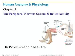
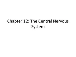
Chapter 12: The Central Nervous System
Chapter 12: The Central Nervous System. Central Nervous System. Brain and Spinal Cord Body’s supercomputer Cephalization – elaboration towards rostal “towards the snout” or anterior portion of CNS Also increase in the number of neurons. Brain. Adult male – 1600 g (3.5 lbs)

Chapter 12: The Central Nervous System
E N D
Presentation Transcript
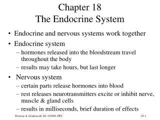
Central Nervous System • Brain and Spinal Cord • Body’s supercomputer • Cephalization – elaboration towards rostal “towards the snout” or anterior portion of CNS • Also increase in the number of neurons
Brain • Adult male – 1600 g (3.5 lbs) • Adult female – 1450 g (3.2 lbs) • Brain mass per body mass - equal
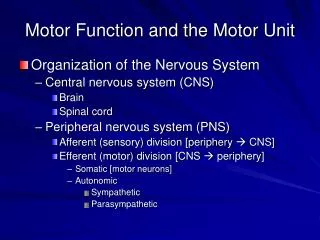
Embryonic Development • 3 week embryo- ectoderm thickens along dorsal midline of body = neural plate • Neural tube invaginates – forms groove flanked by neural folds • Groove deepens – superior ridges fuse forming neural tube – detached from ectoderm and sinks into a deeper position • Neural tube differentiates into CNS – brain anterior and spinal cord - caudal
Embryonic Development 5. Neural crest forms – gives rise to some neurons 6. Neural tube – anterior end expands • 3 primary brain vesicles – • 1. prosencephalon – forebrain • 2. mesencephalon – midbrain • 3. rhombencephalon – hindbrain 7. Week 5 – primary vesicles secondary vesicles • Forebrain telencephalon (endbrain) + diencephalon (hindbrain) • Hindbrain constricts – metencephalon “afterbrain” • Metencephalon – “spinal brain”
Embryonic Development 8. 5 – secondary vesicles – develop into major structures of adult brain • 2 cerebral hemispheres – cerebrum • Diencephalon – hypothalamus • Thalamus • Epithalamus • Retina • Mesencephalon = midbrain • Metencephalon = pons • Myelencephalon = cerebellum • Midbrain and hindbrain = spinal cord
Surfaceectoderm Head Neuralplate Tail The neural plate forms from surface ectoderm. 1 Figure 12.1, step 1
Neural folds Neuralgroove The neural plate invaginates, forming the neuralgroove, flanked by neural folds. 2 Figure 12.1, step 2
Neural crest 3 Neural fold cells migrate to form the neural crest,which will form much of the PNS and many otherstructures. Figure 12.1, step 3
Head Surfaceectoderm Neuraltube Tail 4 The neural groove becomes the neural tube, whichwill form CNS structures. Figure 12.1, step 4
(a) Neural tube (b) Primary brainvesicles Anterior (rostral) Prosencephalon (forebrain) Mesencephalon (midbrain) Rhombencephalon (hindbrain) Posterior (caudal) Figure 12.2a-b
(e) Adultneural canalregions (c) Secondary brainvesicles (d) Adult brainstructures Cerebrum: cerebral hemispheres (cortex, white matter, basal nuclei) Lateral ventricles Telencephalon Diencephalon (thalamus, hypothalamus, epithalamus), retina Third ventricle Diencephalon Cerebral aqueduct Brain stem: midbrain Mesencephalon Metencephalon Brain stem: pons Fourth ventricle Cerebellum Brain stem: medulla oblongata Myelencephalon Central canal Spinal cord Figure 12.2c-e
Regions of Brain • Cerebral Hemispheres • Diencephalon • Brain stem (pons, midbrain, and medulla)
Cortex of gray matter Central cavity Migratory pattern of neurons Inner gray matter Outer white matter Cerebrum Cerebellum Gray matter Region of cerebellum Central cavity Inner gray matter Outer white matter Gray matter Brain stem Central cavity Outer white matter Inner gray matter Spinal cord Figure 12.4
Pattern • Central cavity surrounded by gray matter (neuron cell bodies) • External – white matter (myelinated fiber tracts) • Outer layer of gray matter – cortex – dissapears as you move down the brain stem
Ventricles • Arise from expansions of lumen (cavity) of embryonic neural tube • Filled with cerebral spinal fluid • Lined by ependymal cells
Lateral ventricle Septum pellucidum Anterior horn Posterior horn Inferior horn Interventricular foramen Lateral aperture Median aperture Third ventricle Inferior horn Lateral aperture Cerebral aqueduct Fourth ventricle Central canal (a) Anterior view (b) Left lateral view Figure 12.5
Lateral Ventricles • Deep in cerebral hemisphere • Large C – shaped chambers separated by septum pellucidum • Communicate with 3rd ventricle – in diencephalon • Channel – intraventricular foramen • 3rd ventricle continuous with 4th – canal cerebral aqueduct • 4th ventricle – 3 openings – 2 lateral apertures and median aperture
Lateral ventricle Septum pellucidum Anterior horn Posterior horn Inferior horn Interventricular foramen Lateral aperture Median aperture Third ventricle Inferior horn Lateral aperture Cerebral aqueduct Fourth ventricle Central canal (a) Anterior view (b) Left lateral view Figure 12.5
Cerebral Hemispheres • Superior part of brain • 83 % of brain’s mass • Cover and obscure diencephalon and top of brain stem • Surface – gyri – elevated ridges • Sulci – grooves • Fissures – deeper grooves • Longitudinal fissure – separates cerebral hemispheres • Transverse cerebral fissure – separates cerebral from cerebellum
Cerebral Hemisphere • Divided into 5 lobes by sulci • Central sulcus – frontal plane – separates frontal lobe from parietal lobe • Percentralgyrus and postcentralgyrus – border – central sulcus • Occipital lobe – separate from parietal by parietocciplitalsulcus • Lateral sulcus – outlines temporal lobe • Insula – 5th lobe – deep in lateral sulcus – forms its floor
Precentral gyrus Central sulcus Postcentral gyrus Frontal lobe Parietal lobe Parieto-occipital sulcus (on medial surface of hemisphere) Lateral sulcus Occipital lobe Temporal lobe Transverse cerebral fissure Cerebellum Pons Medulla oblongata Fissure Spinal cord (a deep sulcus) Gyrus Cortex (gray matter) Sulcus White matter (a) Figure 12.6a
Central sulcus Frontal lobe Gyri of insula Temporal lobe (pulled down) (b) Figure 12.6b
Anterior Longitudinal fissure Frontal lobe Cerebral veins and arteries covered by arachnoid mater Parietal lobe Right cerebral hemisphere Left cerebral hemisphere Occipital lobe Posterior (c) Figure 12.6c
Left cerebral hemisphere Transverse cerebral fissure Brain stem Cerebellum (d) Figure 12.6d
Brain • Fits snuggly in skull • Frontal lobes – lie in anterior cranial fossa • Middle cranial fossa – temporal lobe • Each hemisphere – 3 regions 1. cerebral cortex – gray 2. internal white matter 3. basal nuclei
Cerebral Cortex • “executive suite” of NSconscious mind • Enables awareness of ourselves, our sensations, and enables us to communicate, remember, and understand • Also voluntary movement • Composed of gray matter – neuron cell bodies, dendrites, glial and blood vessels
Cerebral Cortex • Billions of neurons – arranged in 6 layers • 2-4 mm thick – 40 % of total brainmass • 52 cortical areas – Broadman areas
Cerebral Cortex • 3 functional areas – • 1. motor area • 2. sensory area • 3. association area • All neurons – interneurons • Each hemisphere – sensory and motor functions of opposite sides of body • Hemispheres – not entirely equal in function • Lateralization or specialization of cortical functions • No functional area acts alone • Conscious behavior involves the entire cortex
Motor areas Sensory areas and related association areas Central sulcus Primary motor cortex Primary somatosensory cortex Premotor cortex Somatic sensation Frontal eye field Somatosensory association cortex Broca’s area (outlined by dashes) Gustatory cortex (in insula) Taste Prefrontal cortex Wernicke’s area (outlined by dashes) Working memory for spatial tasks Executive area for task management Primary visual cortex Working memory for object-recall tasks Vision Visual association area Solving complex, multitask problems Auditory association area Hearing Primary auditory cortex (a) Lateral view, left cerebral hemisphere Motor association cortex Primary sensory cortex Primary motor cortex Sensory association cortex Multimodal association cortex Figure 12.8a
Motor Areas • Control voluntary movement • Posterior part of frontal lobes: primary motor cortex, premotor cortex, Broca’s area, and frontal eye field
Motor Area – Primary Motor Cortex • Primary (somatic) motor cortex – • Located in precentralgyrus of frontal lobe • Large neurons – pyramidal cells • Allow control of precise or skilled voluntary movements • Long axons – project into spinal cord – pyramidal tracts • Somatotrophy – control of body structures mapped to places • Muscles controlled by multiple spots
Posterior Motor Anterior Motor map in precentral gyrus Toes Jaw Primary motor cortex (precentral gyrus) Tongue Swallowing Figure 12.9
Motor Area – Premotor Cortex • Just anterior to precentralgyrus • Controls learned motor skills of repetitious pattern or nature • Coordinates movements of several muscle groups • Memory bank for skilled motor activites
Motor Area – Broca’s Area • Lies anterior to inferior region of premotor area • Considered to be 1. present in one hemisphere only (usually the left) 2. special motor speech area – directs muscles involved in speech production Recently shown to “light up” as we prepare to think or even think about voluntary activities other than speech
Motor Area – Frontal Eye Field • Located partial in and anterior to premotor cortex and superior to Broca’s area • Controls voluntary movement of eyes
Damage • Damage to areas of primary motor cortex – paralyzes the body muscles controlled by those areas • Voluntary control lost, muscles can still contract reflexively • Premotor cortex - damage results in a loss in motor skills programmed in that region, but muscle strength and ability to perform movements are not
Sensory Areas • Occur in parietal lobe, insular, temporal, and occipital lobes 1. Primary Somartosensory Cortex – • In post central gyrus of parietal lobe • Neurons receive info from general (somatic) sensory receptors in the skin and proprioceptors (position sense receptors) in skeletal muscle, joints and tendons • Neurons identify body region being stimulated – spatial discrimination • Right hemisphere – receive input from left side of body • Face & fingertips – most sensitive – largest part
Motor areas Sensory areas and related association areas Central sulcus Primary motor cortex Primary somatosensory cortex Premotor cortex Somatic sensation Frontal eye field Somatosensory association cortex Broca’s area (outlined by dashes) Gustatory cortex (in insula) Taste Prefrontal cortex Wernicke’s area (outlined by dashes) Working memory for spatial tasks Executive area for task management Primary visual cortex Working memory for object-recall tasks Vision Visual association area Solving complex, multitask problems Auditory association area Hearing Primary auditory cortex (a) Lateral view, left cerebral hemisphere Motor association cortex Primary sensory cortex Primary motor cortex Sensory association cortex Multimodal association cortex Figure 12.8a
Sensory Areas 2. Somatosensory Association Cortex – posterior to primary somatosensory cortex • Integrates sensory inputs – temp, pressure, etc. – relayed to produce understanding of object being felt – size, texture, relationship
Posterior Sensory Anterior Sensory map in postcentral gyrus Genitals Primary somato- sensory cortex (postcentral gyrus) Intra- abdominal Figure 12.9
Sensory Areas 3. Visual Areas – primary visual (striate) cortex • Extreme posterior tip of occipital lobe • Most buried deep in calcarinesulcus • Largest • Receives visual input from retina • Visual association areas – surround primary – uses past visual experiences to interpret stimuli
Sensory Areas 4. Auditory Areas – primary auditory cortex – superior margin of temporal lobe • Impulses from ear transmitted here – interpreted as pitch, loudness, location, etc. • Auditory Association area – permits perception of sound • Memories of past sounds
Sensory Areas 5. Olfactory cortex – • Medial aspect of temporal lobes • Small region – piriform lobe • Smell receptors send impulses
Sensory Areas 6. Gustatory Cortex – taste stimuli • Insula just deep to temporal lobe
Sensory Areas 7. Visceral Sensory Area – conscious perception of visceral stimulation • Upset stomach, full bladder, lung bursting – holding breath to long
Sensory Areas 8. Vestibular (equilibrium) cortex – difficult to find • Imaging – shows it in the posterior part of insula and adjacent parietal cortex
Posterior Sensory Anterior Sensory map in postcentral gyrus Genitals Primary somato- sensory cortex (postcentral gyrus) Intra- abdominal Figure 12.9
Multimodal Association Areas • Cortex – complex • Input from multiple senses and outputs to multiple areas • Meaning to info we receive, stores memories, ties to previous experiences, and decide actions • Sensations, thoughts, and emotions
Multimodal Association Areas 1. Anterior Association Areas – frontal lobe – prefrontal cortex • Most complicated • Intellect, complex learning abilities (cognition), recall, and personality • Working memory – abstract ideas, judgment, reasoning, persistence, and planning • Abilities develop slowly in children – region of the brain that matures slowly