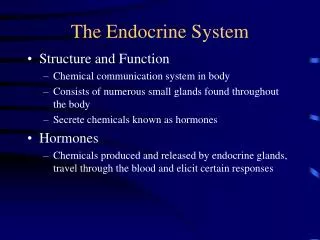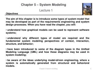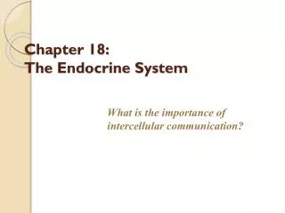
Chapter 18: The Endocrine System
Chapter 18: The Endocrine System. The Nervous and Endocrine Systems…. Act together to coordinate functions of all body systems Nervous system Nerve impulses / Neurotransmitters Faster responses, briefer effects, acts on specific target Endocrine system

Chapter 18: The Endocrine System
E N D
Presentation Transcript
The Nervous and Endocrine Systems… • Act together to coordinate functions of all body systems • Nervous system • Nerve impulses / Neurotransmitters • Faster responses, briefer effects, acts on specific target • Endocrine system • Hormone – a mediator molecule is released in 1 part of the body; but regulates activity of cells in other parts • Slower responses, effects last longer, broader influence Copyright 2009, John Wiley & Sons, Inc.
Endocrine Glands • Two kinds of glands • Exocrine – ducted (sudoriferous, sebaceous, mucous, digestive) • Endocrine – ductless • Secrete products into interstitial fluid, diffuse into blood • Endocrine glands include • Pituitary, thyroid, parathyroid, adrenal and pineal glands • Hypothalamus, thymus, pancreas, ovaries, testes, kidneys, stomach, liver, small intestine, skin, heart, adipose tissue, and placenta - not exclusively endocrine glands Copyright 2009, John Wiley & Sons, Inc.
Hormone Activity • Hormones (H’) affect only specific target tissues with specific receptors http://www.edenatwood.com/ • Receptors constantly synthesized and broken down • Down-regulation = large amounts of H’ leads to a decrease in target-cell receptors • Up-regulation = low amounts of H’ leads to increase in target-cell receptors
Hormone types Circulating – circulate in blood throughout body Local hormones – act locally Paracrine – act on neighboring cells Autocrine – act on the same cell that secreted them
Chemical classes of hormones • Lipid-soluble – use transport proteins produced by ‘liver’ • Steroid H’ - (large diversity) • Thyroid H’ - (iodine added to aa tyrosine) • Nitric oxide - (NO), is both H’ and N’ • Water-soluble – circulate in “free” form • Amine H’ - removal of CO2 (-NH3+) • Peptide H’ (antidiuretic, oxytocin)/ Protein H’ (hGH, insulin) • Eicosanoid H’ (prostaglandins, leukotrienes)
Mechanisms of Hormone Action • Response depends on both hormone and target cell • Lipid-soluble hormones bind to receptors inside target cells • Water-soluble hormones bind to receptors on the plasma membrane • Activates second messenger system • Amplification of original small signal • Responsiveness of target cell depends on • Hormone’s concentration • Abundance of target cell receptors • Influence exerted by other hormones • Permissive (1st activated by another H’), synergistic and antagonistic effects
Lipid-soluble and Water-soluble Hormones Copyright 2009, John Wiley & Sons, Inc.
Blood capillary Blood capillary Blood capillary Blood capillary Free hormone Free hormone Free hormone Free hormone Lipid-soluble hormone diffuses into cell Lipid-soluble hormone diffuses into cell Lipid-soluble hormone diffuses into cell Lipid-soluble hormone diffuses into cell 1 1 1 1 Transport protein Transport protein Transport protein Transport protein 2 2 2 Activated receptor-hormone complex alters gene expression Activated receptor-hormone complex alters gene expression Activated receptor-hormone complex alters gene expression Nucleus Nucleus Nucleus Receptor Receptor Receptor DNA DNA DNA Cytosol Cytosol Cytosol mRNA mRNA mRNA 3 3 Newly formed mRNA directs synthesis of specific proteins on ribosomes Newly formed mRNA directs synthesis of specific proteins on ribosomes Ribosome Ribosome New protein 4 New proteins alter cell's activity Target cell Target cell Target cell Target cell
Blood capillary Blood capillary Blood capillary Blood capillary Blood capillary Blood capillary 1 1 1 1 1 1 Binding of hormone (first messenger) to its receptor activates G protein, which activates adenylate cyclase Binding of hormone (first messenger) to its receptor activates G protein, which activates adenylate cyclase Binding of hormone (first messenger) to its receptor activates G protein, which activates adenylate cyclase Binding of hormone (first messenger) to its receptor activates G protein, which activates adenylate cyclase Binding of hormone (first messenger) to its receptor activates G protein, which activates adenylate cyclase Binding of hormone (first messenger) to its receptor activates G protein, which activates adenylate cyclase Water-soluble hormone Water-soluble hormone Water-soluble hormone Water-soluble hormone Water-soluble hormone Water-soluble hormone Adenylate cyclase Adenylate cyclase Adenylate cyclase Adenylate cyclase Adenylate cyclase Adenylate cyclase Receptor Receptor Receptor Receptor Receptor Receptor Second messenger Second messenger Second messenger Second messenger Second messenger G protein G protein G protein G protein G protein G protein 2 2 2 2 2 ATP ATP ATP ATP ATP Activated adenylate cyclase converts ATP to cAMP Activated adenylate cyclase converts ATP to cAMP Activated adenylate cyclase converts ATP to cAMP Activated adenylate cyclase converts ATP to cAMP Activated adenylate cyclase converts ATP to cAMP cAMP cAMP cAMP cAMP cAMP 6 Phosphodiesterase inactivates cAMP Protein kinases Protein kinases Protein kinases Protein kinases 3 3 3 3 cAMP serves as a second messenger to activate protein kinases cAMP serves as a second messenger to activate protein kinases cAMP serves as a second messenger to activate protein kinases cAMP serves as a second messenger to activate protein kinases Activated protein kinases Activated protein kinases Activated protein kinases Activated protein kinases 4 4 4 Activated protein kinases phosphorylate cellular proteins Activated protein kinases phosphorylate cellular proteins Activated protein kinases phosphorylate cellular proteins Protein Protein Protein ATP ATP ATP ADP ADP ADP Protein— Protein— Protein— P P P 5 5 Millions of phosphorylated proteins cause reactions that produce physiological responses Millions of phosphorylated proteins cause reactions that produce physiological responses Target cell Target cell Target cell Target cell Target cell Target cell
Regulated by Signals from nervous system Chemical changes in the blood Other hormones Most hormonal regulation by negative feedback Few examples of positive feedback Control of Hormone Secretion
Hypothalamus and Pituitary Gland • Hypothalamus is a major link between nervous and endocrine system • Pituitary attached to hypothalamus by infundibulum • Anterior pituitary or adenohypophysis • Posterior pituitary or neurohypophysis Copyright 2009, John Wiley & Sons, Inc.
Hypothalamus and Pituitary Gland Copyright 2009, John Wiley & Sons, Inc.
Anterior pituitary • Release of hormones stimulated by releasing and inhibiting-hormones from the hypothalamus • Also regulated by negative feedback • Hypothalamic hormones made by neurosecretory cells transported by hypophyseal portal system • Anterior pituitary hormones that act on other endocrine systems called tropic hormones Copyright 2009, John Wiley & Sons, Inc.
Hormones of the Anterior Pituitary • Somatotrophs secrete Human growth hormone (hGH) or somatostatin • Stimulates secretion of insulin-like growth factors (IGFs) that promote growth, protein synthesis • Thyrotrophs secrete Thyroid-stimulating hormone (TSH) or thyrotropin • Stimulates synthesis and secretion of thyroid hormones by thyroid • 1. Gonadotrophs secrete Follicle-stimulating hormone (FSH) • Ovaries initiates development of oocytes, testes stimulates testosterone and sperm production • 2. Gonadotrophs secrete Luteinizing hormone (LH) • Ovaries stimulates ovulation, testes stimulates testosterone production
Hormones of the Anterior Pituitary • Lactotrophs secrete Prolactin (PRL) • Promotes milk secretion by mammary glands • Corticotrophs secrete Adrenocorticotropic hormone (ACTH) or corticotropin • Stimulates glucocorticoid (i.e., cortisol) secretion by adrenal cortex • Corticotrophs secrete Melanocyte-stimulating Hormone (MSH) • Unknown role in humans Copyright 2009, John Wiley & Sons, Inc.
Negative Feedback Regulation Copyright 2009, John Wiley & Sons, Inc.
Effects of hGH and IGFs Copyright 2009, John Wiley & Sons, Inc.
1 1 1 1 1 1 1 1 1 1 6 6 6 6 6 Low blood glucose (hypoglycemia) stimulates release of Low blood glucose (hypoglycemia) stimulates release of Low blood glucose (hypoglycemia) stimulates release of Low blood glucose (hypoglycemia) stimulates release of Low blood glucose (hypoglycemia) stimulates release of Low blood glucose (hypoglycemia) stimulates release of Low blood glucose (hypoglycemia) stimulates release of Low blood glucose (hypoglycemia) stimulates release of Low blood glucose (hypoglycemia) stimulates release of Low blood glucose (hypoglycemia) stimulates release of High blood glucose (hyperglycemia) stimulates release of High blood glucose (hyperglycemia) stimulates release of High blood glucose (hyperglycemia) stimulates release of High blood glucose (hyperglycemia) stimulates release of High blood glucose (hyperglycemia) stimulates release of GHRH GHRH GHRH GHRH GHRH GHRH GHRH GHRH GHRH GHRH GHIH GHIH GHIH GHIH GHIH 7 7 7 7 2 2 2 2 2 2 2 2 2 GHRH stimulates secretion of hGH by somatotrophs GHRH stimulates secretion of hGH by somatotrophs GHRH stimulates secretion of hGH by somatotrophs GHRH stimulates secretion of hGH by somatotrophs GHRH stimulates secretion of hGH by somatotrophs GHRH stimulates secretion of hGH by somatotrophs GHRH stimulates secretion of hGH by somatotrophs GHRH stimulates secretion of hGH by somatotrophs GHRH stimulates secretion of hGH by somatotrophs GHIH inhibits secretion of hGH by somatotrophs GHIH inhibits secretion of hGH by somatotrophs GHIH inhibits secretion of hGH by somatotrophs GHIH inhibits secretion of hGH by somatotrophs hGH hGH hGH hGH hGH hGH hGH hGH hGH Anterior pituitary Anterior pituitary Anterior pituitary Anterior pituitary Anterior pituitary 3 3 3 3 3 3 3 3 8 8 8 hGH and IGFs speed up breakdown of liver glycogen into glucose, which enters the blood more rapidly hGH and IGFs speed up breakdown of liver glycogen into glucose, which enters the blood more rapidly hGH and IGFs speed up breakdown of liver glycogen into glucose, which enters the blood more rapidly hGH and IGFs speed up breakdown of liver glycogen into glucose, which enters the blood more rapidly hGH and IGFs speed up breakdown of liver glycogen into glucose, which enters the blood more rapidly hGH and IGFs speed up breakdown of liver glycogen into glucose, which enters the blood more rapidly hGH and IGFs speed up breakdown of liver glycogen into glucose, which enters the blood more rapidly hGH and IGFs speed up breakdown of liver glycogen into glucose, which enters the blood more rapidly A low level of hGH and IGFs decreases the rate of glycogen breakdown in the liver and glucose enters the blood more slowly A low level of hGH and IGFs decreases the rate of glycogen breakdown in the liver and glucose enters the blood more slowly A low level of hGH and IGFs decreases the rate of glycogen breakdown in the liver and glucose enters the blood more slowly 4 4 4 4 4 4 4 9 9 Blood glucose level rises to normal (about 90 mg/100 mL) Blood glucose level rises to normal (about 90 mg/100 mL) Blood glucose level rises to normal (about 90 mg/100 mL) Blood glucose level rises to normal (about 90 mg/100 mL) Blood glucose level rises to normal (about 90 mg/100 mL) Blood glucose level rises to normal (about 90 mg/100 mL) Blood glucose level rises to normal (about 90 mg/100 mL) Blood glucose level falls to normal (about 90 mg/100 mL) Blood glucose level falls to normal (about 90 mg/100 mL) 5 5 5 5 5 5 10 If blood glucose continues to increase, hyperglycemia inhibits release of GHRH If blood glucose continues to increase, hyperglycemia inhibits release of GHRH If blood glucose continues to increase, hyperglycemia inhibits release of GHRH If blood glucose continues to increase, hyperglycemia inhibits release of GHRH If blood glucose continues to increase, hyperglycemia inhibits release of GHRH If blood glucose continues to increase, hyperglycemia inhibits release of GHRH If blood glucose continues to decrease, hypoglycemia inhibits release of GHIH
Posterior pituitary (aka. neurohypohysis) • Does not synthesize hormones • Stores and releases hormones made by the hypothalamus • Transported along hypothalamohypophyseal tract • Oxytocin (OT) • Antidiuretic hormone (ADH) or vasopressin • Key concept: H’ released from PP via nerve impulses Copyright 2009, John Wiley & Sons, Inc.
Hypothalamohypophyseal tract Copyright 2009, John Wiley & Sons, Inc.
Oxytocin (OT) • During and after delivery of baby affects --uterus and breasts • Enhances smooth muscle contraction in wall of uterus • Stimulates milk ejection from mammary glands • In males may foster parental behavior. • Pitocin – synthetic OT Copyright 2009, John Wiley & Sons, Inc.
Decreases urine production by causing the kindeys to return more water to the blood Also decreases water lost through sweating and constriction of arterioles which increases blood pressure (vasopressin) Antidiuretic Hormone (ADH) Copyright 2009, John Wiley & Sons, Inc.
1 1 1 1 1 1 5 5 Low blood osmotic pressure inhibits hypothalamic osmoreceptors Low blood osmotic pressure inhibits hypothalamic osmoreceptors High blood osmotic pressure stimulates hypothalamic osmoreceptors High blood osmotic pressure stimulates hypothalamic osmoreceptors High blood osmotic pressure stimulates hypothalamic osmoreceptors High blood osmotic pressure stimulates hypothalamic osmoreceptors High blood osmotic pressure stimulates hypothalamic osmoreceptors High blood osmotic pressure stimulates hypothalamic osmoreceptors Osmoreceptors Osmoreceptors Osmoreceptors Osmoreceptors Osmoreceptors Osmoreceptors 2 2 2 2 2 Osmoreceptors activate the neurosecretory cells that synthesize and release ADH Osmoreceptors activate the neurosecretory cells that synthesize and release ADH Osmoreceptors activate the neurosecretory cells that synthesize and release ADH Osmoreceptors activate the neurosecretory cells that synthesize and release ADH Osmoreceptors activate the neurosecretory cells that synthesize and release ADH 6 Inhibition of osmo- receptors reduces or stops ADH secretion Hypothalamus Hypothalamus Hypothalamus Hypothalamus Hypothalamus 3 3 3 3 Nerve impulses liberate ADH from axon terminals in the posterior pituitary into the bloodstream Nerve impulses liberate ADH from axon terminals in the posterior pituitary into the bloodstream Nerve impulses liberate ADH from axon terminals in the posterior pituitary into the bloodstream Nerve impulses liberate ADH from axon terminals in the posterior pituitary into the bloodstream ADH ADH ADH ADH Target tissues Target tissues Target tissues 4 4 4 Kidneys retain more water, which decreases urine output Kidneys retain more water, which decreases urine output Kidneys retain more water, which decreases urine output Sudoriferous (sweat) glands decrease water loss by perspiration from the skin Sudoriferous (sweat) glands decrease water loss by perspiration from the skin Sudoriferous (sweat) glands decrease water loss by perspiration from the skin Arterioles constrict, which increases blood pressure Arterioles constrict, which increases blood pressure Arterioles constrict, which increases blood pressure High blood osmotic pressure = • Dehydration • Hemorrhage • Etc.
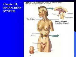
Thyroid Gland • Located inferior to larynx • 2 lobes connected by isthmus • Thyroid follicles produce thyroid hormones • Thyroxine or tetraiodothyronine (T4) • Triiodothyronine (T3) • Both increase (basal metabolic rate) BMR, stimulate protein synthesis, increase use of glucose and fatty acids for ATP production • Parafollicular cells or C cells produce calcitonin • Lowers blood Ca2+ by inhibiting bone resorption Copyright 2009, John Wiley & Sons, Inc.
Thyroid gland Copyright 2009, John Wiley & Sons, Inc.
Thyrotropin-releasing hormone (TRH) from hypothalamus Thyroid-stimulating hormone (TSH) from anterior pituitary Situations that increase ATP demand also increase secretion of thyroid hormones Control of thyroid hormone secretion Copyright 2009, John Wiley & Sons, Inc.
1 1 1 1 1 Low blood levels of T3 and T3 or low metabolic rate stimulate release of Low blood levels of T3 and T3 or low metabolic rate stimulate release of Low blood levels of T3 and T3 or low metabolic rate stimulate release of Low blood levels of T3 and T4 or low metabolic rate stimulate release of Low blood levels of T3 and T3 or low metabolic rate stimulate release of Hypothalamus Hypothalamus Hypothalamus Hypothalamus Hypothalamus TRH TRH TRH TRH TRH 2 2 2 2 TRH, carried by hypophyseal portal veins to anterior pituitary, stimulates release of TSH by thyrotrophs TRH, carried by hypophyseal portal veins to anterior pituitary, stimulates release of TSH by thyrotrophs TRH, carried by hypophyseal portal veins to anterior pituitary, stimulates release of TSH by thyrotrophs TRH, carried by hypophyseal portal veins to anterior pituitary, stimulates release of TSH by thyrotrophs 5 Elevated T3inhibits release of TRH and TSH (negative feedback) TSH TSH TSH TSH Anterior pituitary Anterior pituitary Anterior pituitary Anterior pituitary Anterior pituitary 3 3 3 TSH released into blood stimulates thyroid follicular cells TSH released into blood stimulates thyroid follicular cells TSH released into blood stimulates thyroid follicular cells 4 4 T3 and T4 released into blood by follicular cells T3 and T4 released into blood by follicular cells Thyroid follicle Thyroid follicle Thyroid follicle Actions of Thyroid Hormones: Increase basal metabolic rate Stimulate synthesis of Na+/K+ ATPase Increase body temperature (calorigenic effect) Stimulate protein synthesis Increase the use of glucose and fatty acids for ATP production Stimulate lipolysis Enhance some actions of catecholamines Regulate development and growth of nervous tissue and bones Actions of Thyroid Hormones: Increase basal metabolic rate Stimulate synthesis of Na+/K+ ATPase Increase body temperature (calorigenic effect) Stimulate protein synthesis Increase the use of glucose and fatty acids for ATP production Stimulate lipolysis Enhance some actions of catecholamines Regulate development and growth of nervous tissue and bones Actions of Thyroid Hormones: Increase basal metabolic rate Stimulate synthesis of Na+/K+ ATPase Increase body temperature (calorigenic effect) Stimulate protein synthesis Increase the use of glucose and fatty acids for ATP production Stimulate lipolysis Enhance some actions of catecholamines Regulate development and growth of nervous tissue and bones Actions of Thyroid Hormones: Increase basal metabolic rate Stimulate synthesis of Na+/K+ ATPase Increase body temperature (calorigenic effect) Stimulate protein synthesis Increase the use of glucose and fatty acids for ATP production Stimulate lipolysis Enhance some actions of catecholamines Regulate development and growth of nervous tissue and bones Actions of Thyroid Hormones: Increase basal metabolic rate Stimulate synthesis of Na+/K+ ATPase Increase body temperature (calorigenic effect) Stimulate protein synthesis Increase the use of glucose and fatty acids for ATP production Stimulate lipolysis Enhance some actions of catecholamines Regulate development and growth of nervous tissue and bones
Parathyroid Glands • Embedded in lobes of thyroid gland • Usually 4 • Parathyroid hormone (PTH) or parathormone • Major regulator of calcium, magnesium, and phosphate ions in the blood • Increases number and activity of osteoclasts • Therefore is elevates bone resorption (increase Ca++) • Blood calcium level directly controls secretion of both calcitonin (decrease Ca2+) and PTH via negative feedback Copyright 2009, John Wiley & Sons, Inc.
Roles of Calcitonin, Parathyroid hormone, Calcitrol in Calcium Homeostasis Copyright 2009, John Wiley & Sons, Inc.
1 1 1 1 1 1 3 3 3 3 High level of Ca2+ in blood stimulates thyroid gland parafollicular cells to release more CT. High level of Ca2+ in blood stimulates thyroid gland parafollicular cells to release more CT. High level of Ca2+ in blood stimulates thyroid gland parafollicular cells to release more CT. High level of Ca2+ in blood stimulates thyroid gland parafollicular cells to release more CT. High level of Ca2+ in blood stimulates thyroid gland parafollicular cells to release more CT. High level of Ca2+ in blood stimulates thyroid gland parafollicular cells to release more CT. Low level of Ca2+ in blood stimulates parathyroid gland chief cells to release more PTH. Low level of Ca2+ in blood stimulates parathyroid gland chief cells to release more PTH. Low level of Ca2+ in blood stimulates parathyroid gland chief cells to release more PTH. Low level of Ca2+ in blood stimulates parathyroid gland chief cells to release more PTH. 6 CALCITRIOL stimulates increased absorption of Ca2+ from foods, which increases blood Ca2+ level. 5 5 PTH also stimulates the kidneys to release CALCITRIOL. PTH also stimulates the kidneys to release CALCITRIOL. 4 4 4 2 2 2 2 2 PARATHYROID HORMONE (PTH) promotes release of Ca2+ from bone extracellular matrix into blood and slows loss of Ca2+ in urine, thus increasing blood Ca2+ level. PARATHYROID HORMONE (PTH) promotes release of Ca2+ from bone extracellular matrix into blood and slows loss of Ca2+ in urine, thus increasing blood Ca2+ level. PARATHYROID HORMONE (PTH) promotes release of Ca2+ from bone extracellular matrix into blood and slows loss of Ca2+ in urine, thus increasing blood Ca2+ level. CALCITONIN inhibits osteoclasts, thus decreasing blood Ca2+ level. CALCITONIN inhibits osteoclasts, thus decreasing blood Ca2+ level. CALCITONIN inhibits osteoclasts, thus decreasing blood Ca2+ level. CALCITONIN inhibits osteoclasts, thus decreasing blood Ca2+ level. CALCITONIN inhibits osteoclasts, thus decreasing blood Ca2+ level.
Adrenal Glands • 2 structurally and functionally distinct regions • Adrenal cortex • Mineralocorticoids (aldosterone) affect mineral homeostasis • Glucocorticoids affect glucose homeostasis • Cortisol which increase blood glucose • Androgens have masculinzing effects • Dehydroepiandrosterone (DHEA) only important in females (promote libido) and converted into estrogen • Adrenal medulla • Modified sympathetic ganglion of autonomic nervous system (no axons) • Intensifies sympathetic (fight-or-flight) responses • Epinephrine and norepinephrine Copyright 2009, John Wiley & Sons, Inc.
Adrenal Glands Copyright 2009, John Wiley & Sons, Inc.
Pancreatic Islets • Both exocrine and endocrine gland • Roughly 99% of cells produce digestive enzymes • Pancreatic islets or islets of Langerhans • Alpha or A cells secrete glucagon – raises blood sugar • Beta or B cells secrete insulin – lowers blood sugar • Delta or D cells secrete somatostatin – inhibits both insulin and glucagon • F cells secrete pancreatic polypeptide – inhibits somatostatin, gallbladder contraction, and secretion of pancreatic digestive enzymes Copyright 2009, John Wiley & Sons, Inc.
Pancreas Copyright 2009, John Wiley & Sons, Inc.
Negative Feedback Regulation of Glucagon and Insulin Copyright 2009, John Wiley & Sons, Inc.
1 1 1 1 1 1 1 1 5 5 5 5 Low blood glucose (hypoglycemia) stimulates alpha cells to secrete Low blood glucose (hypoglycemia) stimulates alpha cells to secrete Low blood glucose (hypoglycemia) stimulates alpha cells to secrete Low blood glucose (hypoglycemia) stimulates alpha cells to secrete Low blood glucose (hypoglycemia) stimulates alpha cells to secrete Low blood glucose (hypoglycemia) stimulates alpha cells to secrete Low blood glucose (hypoglycemia) stimulates alpha cells to secrete Low blood glucose (hypoglycemia) stimulates alpha cells to secrete High blood glucose (hyperglycemia) stimulates beta cells to secrete High blood glucose (hyperglycemia) stimulates beta cells to secrete High blood glucose (hyperglycemia) stimulates beta cells to secrete High blood glucose (hyperglycemia) stimulates beta cells to secrete GLUCAGON GLUCAGON GLUCAGON GLUCAGON GLUCAGON GLUCAGON GLUCAGON GLUCAGON INSULIN INSULIN INSULIN INSULIN 2 2 2 2 2 2 2 6 6 6 Glucagon acts on hepatocytes (liver cells) to: Glucagon acts on hepatocytes (liver cells) to: Glucagon acts on hepatocytes (liver cells) to: Glucagon acts on hepatocytes (liver cells) to: Glucagon acts on hepatocytes (liver cells) to: Glucagon acts on hepatocytes (liver cells) to: Glucagon acts on hepatocytes (liver cells) to: Insulin acts on various body cells to: Insulin acts on various body cells to: Insulin acts on various body cells to: • accelerate facilitated diffusion of glucose into cells • speed conversion of glucose into glycogen (glycogenesis) • increase uptake of amino acids and increase protein synthesis • speed synthesis of fatty acids (lipogenesis) • slow glycogenolysis • slow gluconeogenesis • accelerate facilitated diffusion of glucose into cells • speed conversion of glucose into glycogen (glycogenesis) • increase uptake of amino acids and increase protein synthesis • speed synthesis of fatty acids (lipogenesis) • slow glycogenolysis • slow gluconeogenesis • accelerate facilitated diffusion of glucose into cells • speed conversion of glucose into glycogen (glycogenesis) • increase uptake of amino acids and increase protein synthesis • speed synthesis of fatty acids (lipogenesis) • slow glycogenolysis • slow gluconeogenesis • convert glycogen into glucose (glycogenolysis) • form glucose from lactic acid and certain amino acids (gluconeogenesis) • convert glycogen into glucose (glycogenolysis) • form glucose from lactic acid and certain amino acids (gluconeogenesis) • convert glycogen into glucose (glycogenolysis) • form glucose from lactic acid and certain amino acids (gluconeogenesis) • convert glycogen into glucose (glycogenolysis) • form glucose from lactic acid and certain amino acids (gluconeogenesis) • convert glycogen into glucose (glycogenolysis) • form glucose from lactic acid and certain amino acids (gluconeogenesis) • convert glycogen into glucose (glycogenolysis) • form glucose from lactic acid and certain amino acids (gluconeogenesis) • convert glycogen into glucose (glycogenolysis) • form glucose from lactic acid and certain amino acids (gluconeogenesis) 3 3 3 3 3 3 Glucose released by hepatocytes raises blood glucose level to normal Glucose released by hepatocytes raises blood glucose level to normal Glucose released by hepatocytes raises blood glucose level to normal Glucose released by hepatocytes raises blood glucose level to normal Glucose released by hepatocytes raises blood glucose level to normal Glucose released by hepatocytes raises blood glucose level to normal 7 7 Blood glucose level falls Blood glucose level falls 4 4 4 4 4 8 If blood glucose continues to rise, hyperglycemia inhibits release of glucagon If blood glucose continues to rise, hyperglycemia inhibits release of glucagon If blood glucose continues to rise, hyperglycemia inhibits release of glucagon If blood glucose continues to rise, hyperglycemia inhibits release of glucagon If blood glucose continues to rise, hyperglycemia inhibits release of glucagon If blood glucose continues to fall, hypoglycemia inhibits release of insulin
Ovaries and Testes • Gonads – produce gametes and hormones • Ovaries produce 2 estrogens (estradiol and estrone) and progesterone • With FSH and LH regulate menstrual cycle, maintain pregnancy, prepare mammary glands for lactation, maintain female secondary sex characteristics • Inhibin inhibits FSH • Relaxin produced during pregnancy • Testes produce testosterone – regulates sperm production and maintains male secondary sex characteristics • Inhibin inhibits FSH Copyright 2009, John Wiley & Sons, Inc.
Pineal Gland • Attached to roof of 3rd ventricle of brain at midline • Masses of neuroglia and pinealocytes • Melatonin – amine hormone derived from serotonin • Appears to contribute to setting biological clock • More melatonin liberated during darkness than light Copyright 2009, John Wiley & Sons, Inc.
Thymus and Other Endocrine Tissues • Thymus • Located behind sternum between the lungs • Produces thymosin, thymic humoral factor (THF), thymic factor (TF), and thymopoietin • All involved in T cell maturation Copyright 2009, John Wiley & Sons, Inc.
The Stress Response • Eustress in helpful stress / Distress is harmful • Body’s homeostatic mechanisms attempt to counteract stress • Stressful conditions can result in stress response or general adaptation syndrome (GAS) • 3 stages – initial flight-or-fight, slower resistance reaction, eventually exhaustion • Prolonged exposure to cortisol can result in wasting of muscles, suppression of immune system, ulceration of GI tract, and failure of pancreatic beta cells Copyright 2009, John Wiley & Sons, Inc.
Stress Response Copyright 2009, John Wiley & Sons, Inc.
Posterior and Anterior Pituitary Copyright 2009, John Wiley & Sons, Inc.