Download
1 / 43
601 likes | 2.26k Vues
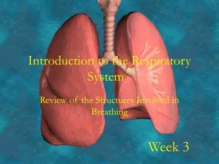
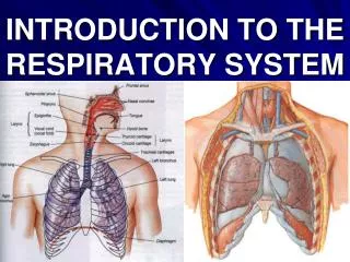
INTRODUCTION TO THE RESPIRATORY SYSTEM. IMPORTANT FACTS:. Oxygen and Carbon Dioxide are respiratory gases Most cells require continuous oxygen supply for production of energy. Without oxygen supply cells die.

E N D
IMPORTANT FACTS: • Oxygen and Carbon Dioxide are respiratory gases • Most cells require continuous oxygen supply for production of energy. • Without oxygen supply cells die. • Carbon dioxide is a waste product of cellular metabolism. Its accumulation leads to acidosis. • Exchange of respiratory gases between cells and their surrounding for homeostasis is maintained by Respiration.
Phylogenesis of Respiratory System • In Protozoa, (unicellular organism) oxygen and carbon dioxide diffuse directly through cell surfaces. • Metazoa(multicellular organisms) developed specific respiratory areas and liquid (blood) transport systems thus providing transportation of respiratory gases . • Mammals have highly developed respiratory and circulatory systems.
RESPIRATORY TERMINOLOGY • Respiration: is a complex process of exchange of respiratory gases (Oxygen & Carbon dioxide) between an organism and external environment. It requires a constant supply of fresh air by Breathing in and out. • Breathing: is a process of continuous ventilation of the lungs when a fresh atmospheric air will replace an air used for the external (pulmonary) respiration • Breathing includes two phases: Inspiration– when air is taken into the lungs and Expiration– when air is removed from these organs • Breathing involves skeletal components of thoracic cage and muscles of respiration, which produce respiratory movements.
Ventilation and Perfusion • During ventilation of lungs a reciprocal movement of air is caused by alternately increasing and decreasing the volume of the chest in breathing. • Perfusion of lungs is achieved via branches of pulmonary arteries, which bring deoxygenated blood to the lungs for its oxygenation
RESPIRATION employs three processes: • External respiration involves the stage of taking oxygen from the air into the blood and returning carbon dioxide to the air. It occurs through the Respiratory Membranes (air-blood barriers). • Transport of respiratory gases in the blood throughout the body via the circulatory system. • Internal or cellular respiration is the process by which glucose or other small molecules within the cell are oxidized to produce energy: this requires oxygen and generates carbon dioxide.
GAS EXCHANGE AT RESPIRATORY MEMBRANE: • Pulmonary ventilation ensures that alveoli are supplied with oxygen, and it removes carbon dioxide arriving from bloodstream. • The actual process of gas exchange occurs between the blood and alveolar air across the respiratory membrane. • The driving force for exchange of respiratory gases is defined by a difference of Partial Pressure of Gases in the air and blood and an ability of their molecules to diffuse from a gas into a liquid and vice versa. • During gas exchange between air & blood molecules of each gas diffuse from area with higher pressure to area with lower pressure (Henry’s Law)
Partial Pressure & Normal Gas Concentration in Air • Inspired air: • Nitrogen – 597 (78.6%) • Oxygen – 159 (20.8%) • Carbon Dioxide – 0.3 (0.04%) • Water Vapor – 3.7 (0.5%) • Alveolar air: • Nitrogen – 573 (75.4%) • Oxygen – 100 (13.2%) • Carbon Dioxide – 40 (5.2%) • Water Vapor – 47 (6.2%) • Expired air: • Nitrogen – 569 (74.8%) • Oxygen – 116 (15.3%) • Carbon Dioxide – 28 (3.7%) • Water Vapor – 47 (6.2%)
Diffusion of Gases-Fick's Law Transfer of gases across cell membranes or capillary walls occurs by simple diffusion. For gases, the rate of transfer by diffusion (X) is directly proportional to the driving force, a diffusion coefficient, and the surface area available for diffusion; it is inversely proportional to the thickness of membrane barrier. Thus, VX- Volume of gas transferred per unit time D- Diffusion coefficient of the gas A- Surface area ΔP- Partial pressure difference of the gas ΔX- Thickness of the membrane
Diffusion of Gases-Fick's Law The driving force for diffusion of a gas is the partial pressure difference of the gas (ΔP) across the membrane, not the concentration difference. Thus, if the PO2 of alveolar air is 100 mm Hg and the PO2 of mixed venous blood that enters the pulmonary capillary is 40 mm Hg, then the partial pressure difference, or driving force, for O2 across the alveolar/pulmonary capillary barrier is 60 mm Hg (100 mm Hg - 40 mm Hg).
Conditions for Oxygenation of Blood: • Sufficient surface areas (Respiratory Areas) for gaseous exchange between Atmospheric air and Blood • Presence of Respiratory Membranes (air-blood barriers) with a very short diffusion path between Atmospheric air and Blood • Concentration gradients for diffusion of Oxygen and Carbon dioxide between the air and blood
Conditions for Oxygenation of Blood: • The surface area available in adult lungs for gaseous exchange is around 140m², which is about the area of a single tennis court. • The blood in the alveolar capillaries is separated from alveolar air by a thin (0.6* in many places) respiratory membrane (1* = one thousandth of a mm).
Respiratory Membrane includes: • 1. Cell membrane & Cytoplasm of the covering alveolar epithelial cell, lined with a layer of Surfactant, which has contact with an air with a high Oxygen content. • 2. Cell membrane & Cytoplasm of endothelial cell of the adjacent blood capillary, containing red blood cells with a high Carbon Dioxide content. • 3. Fused basal laminaeof the above two epithelial cells.
Diffusion gradients are maintained by: • Ventilation (breathing) which renews alveolar air, maintaining oxygen concentration near that of atmospheric air and preventing the accumulation of carbon dioxide. • The flow of blood in alveolar capillaries which continually brings blood with low oxygen concentration and high carbon dioxide concentration to the respiratory membranes.
Sites For External Respiration: • Areas of the body, which may have a presence of Respiratory Membranes with a very short diffusion path between atmospheric air and blood & appropriate concentration gradients of oxygen and carbon dioxide in the air and blood, may perform External Respiration.
Three Types of External Respiration: Lungs, Skin and Alimentary Canal have conditions for External Respiration • Pulmonary respiration - 85%; • Cutaneous respiration - 10%; • Intestinal respiration - 5%.
THREE COMPONENTS OF THE RESPIRATORY SYSTEM: • RESPIRATORY TRACT • Conducting portion • Respiratory portion • LUNGS & PULMONARY CIRCULATION • ADDITIONAL STRUCTURES INVOLVED IN PULMONARY VENTILATION
RESPIRATORY TRACT (AIRWAY) • Its conducting portionis composed of interconnected hollow organs, thus forming a branching out passageway, which has hard elements (bones, cartilages, ligaments) in its walls. Those elements maintain to keep lumina of hollow organs patent, because they prevent collapsing of walls of air passages.
Two Major Subdivisions of Conducting Portion: • a) Upper part/airway (nasal cavity with paranasal air sinuses, oral cavity, nasopharynx, oropharynx and upper portion of laryngopharynx); • b) Lower part/airway (larynx, trachea, extrapulmonary bronchi, intrapulmonary bronchi, regular and terminal bronchioles). It includes bronchial trees.
Upper & Lower Airway Pharynx Nasal Cavity Oral Cavity 4 Larynx 5 6 Trachea
RESPIRATORY TRACT (AIRWAY) • Its respiratory portion – “Respiratory Tree”:consists of functional units “Acini”, which include respiratory bronchioles, alveolar ducts, alveolar atria or vestibules and alveolar sacs with alveoli. Acini are attached to the smallest elements of the bronchial tree – the terminal bronchioles.
Outside air: • Varies in temperature. At the alveolarsurface it must be at body temperature • Varies from very dry to very humid. At the alveolar surface it must be saturated with water vapour • Contains dust and debris. These must not reach the alveolar wall • Contains micro-organisms, which must be filtered out of the inspired air and disposed, before they reach the alveoli, enter the blood and cause possible problems. • It is easy to see that the temperature and humidity of inspired air will increase as it passes down a long series of tubes lined with a moist mucosa at body temperature. The mechanisms for filtering are not so obvious though the turbulence of inspired air could play some role in it.
Functions of Nasal Cavity It is also filtered & cleaned there.
LUNGS • Lungs arepaired parenchymatous organs, which consist of lobes, bronchopulmonary segments and lobules. • They also include intrapulmonary bronchi, bronchioles and numerous Respiratory Trees or functional units: Acini, which contain Respiratory Membranes (air-blood barriers). • Latter serve for gas exchange between atmospheric air and blood. Transport of gases with the blood to and from lungs is provided via the pulmonary circulation.
ADDITIONAL STRUCTURES INVOLVED IN PULMONARY VENTILATION • They include the: • Visceral and parietal pleurae • Two plural cavities (right & left) • Bones and joints of the thoracic cage • Muscles of respiration with their blood and nerve supply)
Mechanism of breathing. • In order to grasp the way in which we breathe we have to grasp the following facts: • Each lung is surrounded by a pleural cavity or sac, except where the plumbing joins it to the rest of the body, rather like a hand in a boxing glove. • The glove has an outer and inner surface, separated by a layer of padding. The pleura, similarly, has two surfaces, but the padding is replaced by a thin layer of fluid. • Each lung is enclosed in a cage bounded below by the diaphragm and at the sides by the chest wall and the mediastinum. • Breathing works by making the cage bigger: the pleural layers slide over each other and the pressure in the lung is decreased, so air is sucked in. Breathing out does the reverse, the cage collapses and air is expelled.
Ribs, Respiratory Muscles, Lungs & Pleurae Visceral Pleura Parietal Pleura
Mechanism of breathing. • Breathing movements are sometimes divided into: • Pump handle movements, the sternal end of rib is elevated or depressed on its vertebral joints • Bucket handle movements, the rib rotating on its axis around anterior and posterior attachments. • With more and more effort put into deeper and deeper breathing the scalene muscles of the neck contract, raising the first rib and hence the rest of the cage, then other neck muscles and even those of the upper limb become involved. • A patient with difficulty in breathing often grips a table edge in order to stabilize the limbs so that their muscles can be used to help in moving the thoracic wall.