

SlideServe
Menu
Préférences
© SlideServe · v2026

MALDI- TOF

mprasadnaidu avatar
19 diapositives · 1.5K Views · Feb 11, 2015

BIOCHEMISTRY

MALDI- TOF - slide 1
01 / 19MALDI- TOF
1 / 17

Télécharger la présentation

MALDI- TOF
Une image / un lien ci-dessous est fourni (tel quel) pour télécharger la présentation

Politique de téléchargement : le contenu du site vous est fourni TEL QUEL pour votre information et votre usage personnel ; il ne peut être vendu, concédé sous licence ni partagé sur d’autres sites sans l’accord de son auteur.Le contenu vous est fourni TEL QUEL, pour votre information et votre usage personnel uniquement. Téléchargez la présentation en cliquant sur ce lien. Lors du téléchargement, si vous ne parvenez pas à télécharger une présentation, l’éditeur a peut-être supprimé le fichier de son serveur.Pendant le téléchargement, si vous ne parvenez pas à obtenir une présentation, le fichier a peut-être été supprimé par l’éditeur.

F I N

Plus de contenu associé

Transcription de la présentation
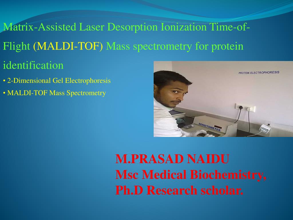
  1. Matrix-Assisted Laser Desorption Ionization Time-of-Flight (MALDI-TOF) Mass spectrometry for protein identification • 2-Dimensional Gel Electrophoresis • MALDI-TOF Mass Spectrometry M.PRASAD NAIDU Msc Medical Biochemistry, Ph.D Research scholar.

  2. The age of X-omics and biotechnology: • Genomics: Human genome project • Transcriptomics: cDNA microarray • Proteomics: • Development and involvement of mass spectrometry Celera Genomics Inc. cDNA microarray Tandem mass spectrometer (MS/MS) MALDI-TOF MS

  3. Proteomics solution IEF SDS-PAGE

  4. 2-Dimension Electrophoresis (2-DE) for Protein Separation The core technology of proteomics is 2-DE: At present, there is no other technique which is capable of resolving thousands of proteins in one separation procedure. Speaker: C. C. Wu Date: 31/10/2001

  5. NH3+ NH3+ NH2 COOH COO- COO- NH3+ NH3+ NH2 COOH COO- COO- pH < pI Positive charge pH = pI pH > pI Negative charge Isoelectric point Net charge pH Isoelectric point (pI): Isoelectric point is the pH of a solution at which the net charge of protein is zero. In electrophoresis there is no motion of the particles in an electric field at the isoelectric point.

  6. General principle and protocol of 2-Dimension Electrophoresis Ampholytes sample Isoelectric focusing (1st dimension) pH 9 - pH 3 + polyacrylamide 2nd dimension SDS-PAGE MW pH gradient

  7. Traditional Equipment for Isoelectric focusing (IEF): Ampholytes polyacrylamide Cathode (-) electrode solution Anode (+) electrode solution

  8. Traditional 2-Dimensional Electrophoresis Disadvantage: cathodic drift Cathode (-) electrode solution pH 9 pH 7 pH 5 Ampholyte polyacrylamide pH 3 pH 3 pH 3 Time Anode (+) electrode solution

  9. CH2 = CH-C-NH-R O Immobilized pH Gradient (IPG) Acrylamide monomer Acidic buffering group: COO- Basic buffering group: NH3+ Polyacrylamide gel

  10. acidic basic Production of Immobilized pH Gradient (IPG) strip Gradient maker A D plastic support film B E C F pH 3 pH 10

  11. Equipment for Isoelectric focusing (IEF): IPGphor (IEF System) Amersham Pharmacia Biotech Inc. Protein IEF Cell Bio-Rad Laboratories

  12. Sample preparation Lysis solution: 8M Urea 4% NP-40 or CHAPS 40mM Tris base Cell line Lysis solution Sonication vacuum Lysis solution Centrifugation Measurement of [protein] 2-DE sample

  13. IPG strip rehydration and sample loading Rehydration solution 2-DE sample Rehydration solution: 8M Urea 2% NP-40 or CHAPS 2% IPG buffer (Ampholyte) 0.28% DTT Trace Bromophenol blue IPG strip holder Position the IPG strip

  14. IPG strip rehydration and sample loading Strip holder Anode (+) electrode Cathode (-) electrode 30 voltage 12hr

  15. Electrode pads Holder cover IPG strip Electrode Voltage Time First dimension: Isoelectric focusing 1. Place electrode pads (?) 2. 200 V step-n-hold 1.5hr 3. 500 V step-n-hold 1.5hr 4. 1000 V gradient 1500vhr 5. 8000 V gradient (?) 36000vhr

  16. SDS SDS-PAGE SDS-PAGE Marker in paper 0.5% agarose in running buffer SDS-PAGE • Second dimension: SDS-PAGE • SDS equilibration • SDS-PAGE SDS equilibration buffer 50 mM Tris-HCl 6 M Urea 30% Glycerol 2% SDS Trace Bromophenol IPG strip

  17. Protocol of silver stain: 50% methanol 25% acetic acid 4hr ddH2O 30 sec ddH2O x 3 times 30min/time 3% Na2CO3 0.0185% formaldehyde 0.004% DTT solution 30min 2.3M citric acid 0.1% AgNO3 30min 5% acetic acid 25% methanol

  18. 2-DE separation of soluble E. coli proteins

  19. THANK YOU