Introduction to MALDI-TOF
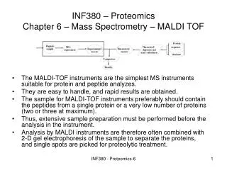
Introduction to MALDI-TOF. Features of MALDI-TOF MS. Soft ionization - analyze intact biomolecules and synthetic polymers Broad mass range - analyze a wide variety of biomolecules Simple mixtures are okay Relatively tolerant of buffers and salts Fast data acquisition


Introduction to MALDI-TOF
E N D
Presentation Transcript
Features of MALDI-TOF MS • Soft ionization - analyze intact biomolecules and synthetic polymers • Broad mass range - analyze a wide variety of biomolecules • Simple mixtures are okay • Relatively tolerant of buffers and salts • Fast data acquisition • Easy to use and maintain, no water or gas hook ups required • High sensitivity, superior mass resolution and accuracy
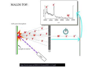
MALDI: Matrix Assisted Laser Desorption Ionization Laser Sample plate hn • 1. Sample (A) is mixed with excess matrix (M) and dried • on a MALDI plate. • 2. Laser flash ionizes matrix molecules. • 3. Sample molecules are ionized by proton transfer from matrix: • MH+ + A M + AH+. AH+ Variable Ground Grid Grid +20 kV
Time-of-flight mass analyzer Source Drift region (flight tube) + + detector + + V • Ions are formed in pulses. • Small ions reach the detector before large ones. • Measures the time for ions to reach the detector.
Calibration of the mass scale The mass-to-charge ratio of an ion is proportional to the square of its drift time. t = Drift time L = Drift length m = Mass K = Kinetic energy of ion z = Number of charges on ion
Voyager-DE STR MALDI TOF Sample Linear Extraction plate Reflector detector grids Timed ion detector Attenuator selector Reflector Prism Laser Collision cell Camera Pumping Pumping
MALDI TOF Hardware Laser, Attenuator and Prism Nitrogen laser at 337 nm, 3 ns wide pulses, 20 Hz. Laser attenuator varies the intensity of the laser hitting the sample. Prism deflects the laser beam into the ion source. Sample Plate and Sample Stage An accelerating voltage is applied to the sample plate in the range 15-25 kV. Variable Voltage Grid A grid 1-2 mm above the sample plate with an additional voltage to fine- tune ion acceleration Ground Grid Grounded surface defines end of acceleration region Grounded Aperture Entrance to flight tube
MALDI TOF Hardware Vacuum System High vacuum is required to avoid ion collisions Flight tube A field free region where ions drift at a velocity inversely proportional to the square root of their mass/charge. Linear Detector Measures the ion abundance in linear mode (no reflector used) and sends a signal to the digitizer.
Ions are detected with a microchannel plate primary ion - 1000V + - e - e L - e - e - 100V D L >> D
High current detector schematic Used in linear mode to enhance signal from high mass molecules Fast scintillator Microchannel plate Condenser Photomultiplier tube signal 1kV 15kV
MALDI TOF Hardware Reflector A single stage gridded ion mirror that subjects the ions to a uniform repulsive electric field to reflect them. It is tilted by 1° in the DE-STR to focus the ions on to the detector Collision Cell Gas cell for collision induced dissociation (CID) to enhance fragmentation in PSD analysis Reflector Detector Measures ions reflected by the mirror. In the DE-STR this is a 6-10 mm pore size micro-channel plate. Timed Ion Selector A velocity selector that allows a single precursor ion of a selected mass and their fragment ions to pass to the detector. A Bradbury-Neilson gate is used.
Voyager-DE STR MALDI TOF Sample Linear Extraction plate Reflector detector grids Timed ion detector Attenuator selector Reflector Prism Laser Collision cell Camera Pumping Pumping
The problem: Peaks are inherently broad in MALDI-TOF spectra (poor mass resolution). The cause: Ions of the same mass coming from the target have different speeds. This is due to uneven energy distribution when the ions are formed by the laser pulse. Sample + matrix on target Ions of same mass, different velocities + + +
Can we compensate for the initial energy spread of ions of the same mass to produce narrower peaks? Delayed Extraction Reflector TOF Mass Analyzer
Delayed Extraction (DE) improves performance Ions of same mass, different velocities 0 V. + + + 0 V. Step 1: No applied electric field. Ions spread out. + 20 kV. + + 0 V. Step 2: Field applied. Slow ions accelerated more than fast ones. + 20 kV. + + 0 V. Step 3: Slow ions catch up with faster ones.
What is a reflector TOF analyzer? A single stage gridded ion mirror that subjects the ions to a uniform repulsive electric field to reflect them. Detector Ion Source Reflector (Ion Mirror) The reflector or ion mirror compensates for the initial energy spread of ions of the same mass coming from the ion source, and improves resolution.
A reflector focuses ions to give better mass resolution + + 0 V. +20 kV
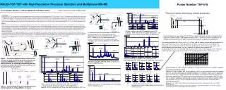
Resolution = 18100 8000 15 ppm error 6000 Resolution = 14200 4000 Counts 24 ppm error Resolution = 4500 2000 0 55 ppm error 2840 2845 2850 2855 Mass (m/z) Resolution & mass accuracy on mellitin
Fundamentals of Post Source Decay(PSD) • PSD refers to a method of detecting and measuring the masses of fragment ions that are formed from a selected precursor ion. • Fragment ions are mainly formed by unimolecular decomposition after the precursor ions are fully accelerated (after they exit the source—hence post-source decay) • Fragment ions are separated and detected in the reflector.
Decomposition occurs in the flight tube Lineardetector Reflector detector Laser Decay can occur at any point along here Reflector Source
Internal energy of precursor ions No of ions Internal energy Only a small fraction of the precursor ions have enough energy to fragment during their lifetimes. For peptides the efficiency of PSD fragmentation is amino acid composition and sequence dependent.
Increasing PSD Fragmentation • There are two ways to increase the amount of fragmentation: both act to increase the precursor ions’ internal energy. • Use higher laser intensity • Use a collision cell
+ + + PSD fragment ion velocities are the same as their precursors All three of these species travel at the same velocity in the flight tube until they reach the reflector. Why? Velocity is determined by initial acceleration. Initial energy = 20 keV. Bond energies = ~ 10 eV, so breaking a bond has a very minor effect on velocities.
Timed Ion Selector (TIS) The TIS is a Bradbury-Neilson gate, which is a type of velocity selector. It allows only selected precursor ions and their fragments to pass through to the reflector. Gate closed: alternating potentials on wires Gate open: wires at ground potential + - Ions + -
Timed Ion Selector operation TIS off “Gate open” TIS on “Gate closed”
Before fragmentation The intact molecular ion has translational kinetic energy equal to: KE = 1/2 Mv2 where: KE = kinetic energy (= zeV) M = mass v = velocity
Post source fragmentation The translational kinetic energy of a fragment ion is where KEM = precursor kinetic energy KEm = fragment kinetic energy M = precursor mass m = fragment mass
+ + Precusor and PSD fragment ions take different paths in the “normal” reflector Reflectordetector Intact precursor ion Fragment ion formed by PSD 0 V. +20 kV Reflector
How are PSD fragment ions that are traveling at the same speed as the precursor ion but contain reduced kinetic energy made to arrive at the detector so that they are focused? By varying the “steepness” of the voltage gradient in the reflector across the fragment ion mass range.
MH+ AH+ + B MH+ A + BH+ PSD mirror ratio setting Consider an ion (MH+) that can decompose into two fragments, A and B. Either of the following reactions can occur: Assume MH+ = 1,000 Da, AH+ = 700 Da, and BH+ = 300 Da
At mirror ratio = 1.00 MH+ ( 1,000) correctly focused AH+ (700) Poorly focused BH+ (300) Poorly focused AH+ BH+ MH+
At mirror ratio = 0.7 BH+ AH+ MH+ MH+ ( 1,000) not focused AH+ (700) correctly focused BH+ (300) Poorly focused
At mirror ratio = 0.3 BH+ AH+ & MH+ MH+ ( 1,000) not focused AH+ (700) not focused BH+ (300) correctly focused
Resolution decreases as the fragment ions penetrate less into the mirror MR=0.80 MR=0.71 MR=0.61 (MR= mirror ratio)
PSD Spectrum of Angiotensin I, MH+ = 1296.7 Da Composite of the focused mass regions from several spectra acquired with different mirror ratios
Characteristics of CID (collison induced dissociation) • Immonium ion signals are enhanced with collision gas; use routinely below fragment mass 200. • Collisions can induce fragmentation of ions that do not decompose under normal PSD conditions. • Side chain fragmentation may allow one to distinguish between Leu and Ile.
CID to distinguish between Ile and Leu (GRF Lys-C peptide KLLQDILSR; MH+ = 1085.667)