
SPECTROPHOTOMETRY
E N D
Presentation Transcript
SPECTROPHOTOMETRY M.PRASAD NAIDU MSC MEDICAL BIOCHEMISTRY
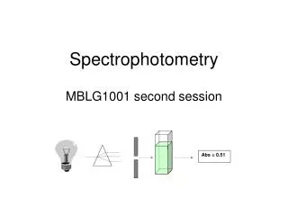
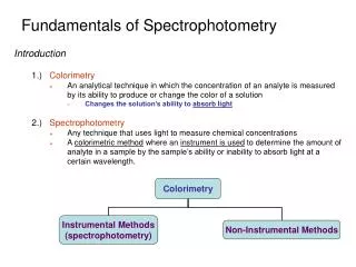
SPECTROPHOTOMETRY :- Defined as the measurement of intensity of light at selected wave length .
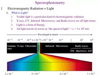
Basic principles of light Light has dual characteristics Photon ( energy ) Wave form
Photon Energy packets . E = h v h = Planck’s constant ( 6.22 x 1027erg sec ) Frequency ( v ) Number of wave passing through a fixed point per second . v = c / λ c = speed of light in vaccum (3x10¹⁰cms/sec ) Wave length (λ ) Distance between two peaks as the light travels in wave like manner . Expressed in nanometers ( nm ).
Relationship b/n Transmittance , Absorbance Transmittance ( T ) =Is / Io % T = Is / Io x 100 A = - log10 T O D = -log10 % T
The Laws of Absorption 1. Beer s Law :- States that concentration of a substance is directly proportional to the amount of light absorbed or inversely proportional to the logarithm of the transmitted light
2. Lambert s Law :- States that the amount of light absorbed is proportion to the thickness of absorbing material and is independent of the intensity of the incident light .
A = abc A = Absorbance a = proportionality constant (absorptivity) b = Light path in cms c = concentration of the absorbing compound in g/L
Light Source • Incandescent :- visible spectrum------Tungsten light bulb uv spectrum --------Hydrogen & deuterium lamps atomic absorption-----Hollow cathode lamp • Laser sources :- These provide intense light and narrow wave length .
Monochromater ( Spectral isolation ) System for isolating radiant energy of a desired wave length . • Filters • Prisms • Diffraction gratings Slits may be inserted before & after the monochromater device to render light rays parallel or to isolate narrow portion of the light beam .
Filters : - • simplest • Thin layer of colored glass • Operates by absorbing light in all other region except for one ,which they reflect . • Resolve polychromatic light into a relatively wide band width ( 50 nm ) • Used only in colorimeter Disadvantage • Low transmittance ( 5 – 20 % ) Normal T = 20 -80 % A = 0.1 -0.7
The color of filter should be complementary to the color of the solution
Prisms :- • Separates white light into a continues spectrum by refraction ----shorter wave length are refracted more than longer wave length . • this results in non linear with the longer wave length closer together .
Diffraction gratings:- • Prepared by depositing a thin layer of aluminium –copper alloy on the surface of a flat glass plate .Then ruling many parallel grooves into the metal coating . • These are then used as moulds to prepare less expensive replicas for instrumental use . • Better gratings ------1000—2000 lines /mm .
Cuvetts • Absorption cells • Shapes ---round square rectangle • Material ---glass silica (quartz ) plastic • All have constant path length ---1cm
Precautions • Cuvetts must be clean & optically clear • Etching / deposition on the surface effects absorbance value • Cuvetts are cleaned by copious rinsing with distilled water • Wash with mild detergent or soak in a mixture containing HCl :H2O: Ethanol ( 1: 3 : 4 )
Cont---- • Alkaline solution not left standing for prolonged period as it dissolves glass and produces etching • Never soak in dichromate cleaning solution as it is hazardous and tends to adsorb onto and discolor the glass • Invisible scratches , finger prints or residual traces of previously measured substance may interfere with absorbance ( uv-vis spectrophotometry )
Cont d --- A good practice is to fill all Cuvetts with distilled water and measure the absorbance for each against a reference blank over the wavelength to be used . This value should be essentially ZERO
Photo detectors Converts light into an electric signal that is proportional to the number of photons striking its photosensitive surface . Commonly used are • Photomultiplier tube • Solid state detectors -Photodiodes -Charge couple detectors
Read out devices Electrical energy from a detector is displayed on meter or read out system . • Direct reading system no further amplification . • Digital read out device Provides visual numerical display of absorbance or converted values of concentration
Performance parameters To verify that a spectrophotometer is performing satisfactorily or not . Parameters tested • Wave length accuracy • Special band width • Stray light • Linearity • Photometric accuracy
Deviation from Beer Lambert s Law Reasons -High sample concentration Specimens may polymerize or ionize Coagulate to form turbid solution ( higher absorbance ) -Instrumentation limitations Imperfect monochromacy Stray lights Power fluctuations
Temperature effects Changes in temp changes the degree of solubility ,dissociation /association properties of the solute ,hydration etc. So absorbance measurement must always be done at constant temp . • Sample instability Color developed may be unstable
Application of UV-VIS Spectrophotometer 1. Qualitative analysis -identify compounds in pure state \in biological preparation -by plotting a absorption graph -curves are specific to a compound eg:- - Nucleic acid 254 nm -Proteins 280 nm
Absorption of compounds in different region gives some hint of its structure 220 – 280nm No absorption aliphatic – alicyclic hydrocarbons 220 -250nm absorption + two unsaturated linkages Benzidine derivative 250 – 300nm absorption + more than two double bonds
2 .Quantitative analysis: comparing the absorbance of a sample (unknown concentration) with standard (with known concentration)
3. Enzyme assay: Determination of enzyme activity – change in absorbance Eg; LDH Lactate + NAD+ Pyruvate + NADH+H+ ( 340nm) Coupled enzyme assay: Eg; PK PEP + ADP Pyruvate + ATP LDH Pyruvate + NADH+H+ Lactate + NAD+ ( 340nm)
4. Study of cis –trans isomerism: • Differs in spatial arrangement of groups around the plane. So absorption spectra also differs • Trans ------- more elongated -----maximum absorption at longer wave length.
5 .Control of purification: • Impurities in a compound easily detected Eg; carbon disulphide in carbon tetrachloride (impurity, 318nm) Benzene impurity in commercial alcohol (280nm) (210nm)