Download
1 / 33
340 likes | 881 Vues
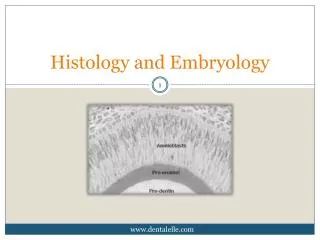
HISTOLOGY & EMBRYOLOGY Teaching PPT Dept. of Anat., Hist. & Embry. School of Medicine Xi’an Jiaotong University. Male Reproductive System. Qiu Shudong. Introduction. Testes Genital ducts: Epididymis, Ductus deferens, Ejaculatory ducts Urethra Accessory glands:

E N D
HISTOLOGY & EMBRYOLOGY Teaching PPT Dept. of Anat., Hist. & Embry. School of Medicine Xi’an Jiaotong University
Male Reproductive System Qiu Shudong
Introduction Testes Genital ducts: Epididymis, Ductus deferens, Ejaculatory ducts Urethra Accessory glands: Seminal vesicles, Prostate, Bulbourethral gland Penis
TESTES I. General Structure : Tunica vaginalis (mesothelium) Testicular Tunica albuginea & capsule mediastinum testis Tunica vasculosa (l.c.t & Cap.) Septula testis Testicular Seminiferous tubule lobules Straight tubules,rete testis & efferent ductules Interstitial tissue & Leydig cell Function: Produce sperm & sexual hormone
Seminiferous tubule Interstitial tissue Testicular Capsule Straight tubule Testicular lobules
II. Seminiferous Tubules D = 150 - 250um, 30 - 70cm long, Spermatogenic Spermatogenic cells epithelium Sertoli cells Basement membrane Collagenous fibers Limiting membrane Myoid cells
Spermatogenic epithelium Limiting m. Sertoli Cell Interstitial tissue & Leydig cell
(l) Spermatogenic cell & Spermatogenesis • At puberty / FSH stimulating • Spermatogenic cells differentiate to spermatozoa • Successive spermatogenic cells : • spermatogonia • primary spermatocyte • secondary spermatocyte • spermatids • spermatozoa. • Spermatogenesis: The process from spermatogonia to spermatozoa. • Including 3 phases:
A. Spermatocytogenesis: spermatogonia primary spermatocytes by mitosis B. Meiosis: Spermatocytes spermatids, through 2 succeesive meiosis w/ reduction by half number of chromosomes & half amount of DNA per cell C. Spermiogenesis: Spermatids spermatozoa by modifying structures & shape (no division).
Spermatocytogenesis primary Meiosis secondary spermatids Spermiogenesis spermatozoa
Spermatogenesis& Spermiogenisis Primary Spermatocyte 1st meiosis 2nd meiosis Secondary Spermatocyte Spermatid Spermatozoon
(2) Spermatogonia & Spermatocytogenesis • Spermatogonium: The most immature spermatogenic cell, on B.M. Round & small (12 um), a round nucleus w/ l-2 nucleoli & finely-granulated chromatin. • Spermatocytogenesis: At sexual maturity, a series of successive mitosesto form: (l) spermatogonia A (stem cells); (2) spermatogonia B, ( primary spermatocyte).
46 chromasomes & 2n DNA 46 chromasomes & 2n DNA 46 chromasomes & 4n DNA
(3) Primary & 2nd spermatocyte & meiosis: • Primary spermatocyte In middle zone of the epithelium. Easy to seen in section (the first meiosis need 22 days) The largest one (16-18um) , nucleus w/ prominent chromosomes, 46 (44 +XY) & 4N DNA. Primary spermatocyte →→→ Secondary spermatocyte(by 1st meiosis) • Secondary spermatocyte Near lumen. Difficult to observe in section (the second meiosis quickly completed). Round (12 um) & a pale-stained nucleus, 23 ( 22+X or 22+y) & 2N DNA. Secondary spermatocyte →→→ spermatids(by 2nd meiosis)
46 chromasomes& 2n DNA 46 chromasomes& 4n DNA 23 chromasomes& 2n DNA 23 chromasomes & 1n DNA
(4) Spermatid & spermiogenesis • Spermatid Close to lumen. spherical (6 um). 23 (22+X or 22+Y) & 1N DNA (haploid) • Spermiogenesis A complex process that a round spermatid transforms into a tadpole-like spermatozoon (no further division occurs) . Including:
(a) Golgi apparatus acrosomal granule acrosomal vesicle acrosomal cap acrosome (covers 2/3 of the nucleus anteriorly) (b) Condensation of chromatin a elongated & flattened nucleus. (c) A centriole a flagellum (axoneme) At the posterior pole of the nucleus ( for motility). (d) Mitochondria a sheath of mitochondria. Spiral around initial part of the tail (e) Parts of the cytoplasm cast off as a residual bodies (phagocytized by Sertoli cells). (a) Golgi apparatus acrosomal granule acrosomal vesicle acrosomal cap acrosome (covers 2/3 of the nucleus anteriorly) (b) Condensation of chromatin a elongated & flattened nucleus. (c) A centriole a flagellum (axoneme) At the posterior pole of the nucleus ( for motility). (d) Mitochondria a sheath of mitochondria. Spiral around initial part of the tail (e) unnecessary cytoplasm cast off as a residual bodies (phagocytized by Sertoli cells).
(5) Human Spermatozoon: • 60 um long, consist of a head & a tail. • Head: pear-shaped & flattened w/ a nuclear & a acrosome (contains hydrolytic enzyme important for fertilization). • Tail: 55um in length w/ a microtubular axoneme in the core. Subdivided into 4 segments: (a)The neck, containing a centriole (connect) (b)The middle segment (5-7um), containing a sheath of mitochondria (provide energy). (c)The principal segment (45um), containing a fibrous sheath (support the tail). (d)The end piece (5-7um), containing a microtubular axoneme.
Mis. Egg , I miss you very much! Mr. sperm, Mr. Sperm , I miss you very much too !
The Passage of the Sperm Seminiferous tubules straight tubules rete testis efferent ductules genital ducts
2. Sertoli cells: Columnar cell rest on B.M., the free surface reaches to lumen. spermatogenic cells locate b/w adjacent cells Features & function: LM: no clear outline, basally-located, ovoid nucleus w/ a distinct nucleolus. EM: (l) abundant sER., Gl., Ly., Mf., & Mt. (2) Tight junctions present b/w adjacent Sertoli cells (isolate spermatogonia from other spermatogenic cells).
S E R T O L I C E L L
C. Function: (l) Support & nourish spermatogenic cells; (2) Secrete fluid to help the sperm moving; (3) Phagocytize & digest the residual bodies (4) Synthesize & secrete ABP (androgen binding protein) which combines androgen in seminiferous tubule to stimulate spermatogenesis; (5) Form the blood-testis barrier: Tight junctlon constitute the main part (rest: B.M. & limiting membrane). Fnction: separates germ cells from immune system & prevents auto-immune reaction. (6) Preventsome physical & chemical factors from damaging germ cells, e.g. radiation, body temperature, infection
III. Interstitial tissue • B/w seminifeious tubules • L.c.t. w/ small b.v. & l. v., nerves & Leydig cells: a. in groups & near b.v.; b. large, round or polygonal, acidophilic, round nucleus w/ prominent nucleolus. c. EM: abundant sER, tubular cristae Mit., lipid droplets; *rod-shaped crystalloids.
Function: secrete testosterone • for proliferation & differentiation of germ cells, • for development of genital system, • for maintenance of male secondary sex characteristics.
IV. Regulation of testicular function: 1. Leydig cells are controlled by interstitial cell stimulating hormone (ICSH); 2. Follicle stimulating hormone (FSH) regulates the function of Sertoli cells 3. ICSH & FSH are secreted by cells in glandular pituitary.
Epididymis I. A head, a body & a tail. II. Consist of a highly coiled ductus epididymis iii. The ductus is lined a pseudostratified columnar epithelium • The principal cells have numerous stereocilia • The base cells may be germinative
IV. Function: • Absorb most of the fluid that leaves the testis • Secrete carnitine, glycerylphosphoryo-choline, sialic acid, etc. • Spermatozoa become mature functionally, acquiring motility & fertilizability when they slowly pass the ductus
prostate • Consist of compound glands around the urethra, c.t. and smooth muscles • Glandular epithelium is columnar, cuboidal or pseudostratified. • Prostatic concretions may be found in the alveoli • Function: The secretion containing acid phosphatase makes up the main part of seminal plasma * Benign hypertrophy of prostate can lead to obstruction of the urethra. ** Prostate cancer developes in main glands