Download

1 / 15
150 likes | 283 Vues
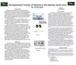
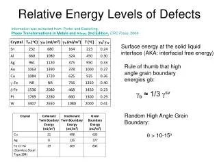
A Comparison of the Relative Actin Levels in Various Organs in Danio rerio Embryos. Mari Ann Elise Springer Michael Raymond Bukszar Elizabeth Rae Kellom. Introduction. Actin. Essential for cell movement Dynamic Monomers vs. filaments Found in all cells but especially crucial in muscle.

E N D
A Comparison of the Relative Actin Levels in Various Organs in DaniorerioEmbryos Mari Ann Elise Springer Michael Raymond Bukszar Elizabeth Rae Kellom
Introduction Actin • Essential for cell movement • Dynamic • Monomers vs. filaments • Found in all cells but especially crucial in muscle
Introduction Organs of Focus • Skeletal Muscle • Heart • Brain 48hr
Introduction Striated vs. Cardiac Muscle
Hypothesis • Actin levels in the organs of zebrafish embryos are differentiated based on muscle density showing actin levels being highest in skeletal muscle, moderate in the heart, and lowest in areas with minimal muscle such as the brain.
Methods Techniques • Rhodamine Phalloidin staining • Permeabilization
Methods Methods (Halpern & Gamse, 2009) • 10 3-day-old zebrafish embryos fixed in 4% formaldehyde • Washed in PBS + .1% Tween • Permeabilizedin PBS + 2%Triton for 2 hours • Washed in PBS tween • Incubated with phalloidinfor staining; kept in dark from this point onward • Washed in PBS tween • Glycerol Transfer series: 25%, 50%, 80% • Embryos were examined and photographed under a fluorescent microscope; 50 millisecond exposure time • Ear and eye analyzed instead of brain • Pictures were analyzed for relative actin levels using ImageJ and compared them using One Way ANOVA
Results Heart Eye Otic Vesicle Skeletal Muscle
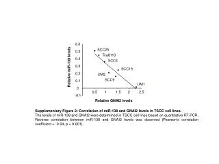
Results Figure 1. Relative actin levels in respective Danioriero organs
Results Figure 2. One-way ANOVA of actin levels between organs in Danioriero
Discussion Relation to Hypothesis • Hypothesis changed slightly during the experiment • Results did not support hypothesis • Few suitable fish for observation & analysis
Discussion Dynamic Hypothesis 2-day: 5-day:
Discussion Reasoning • Extraocular muscles are small & deep • Actin is important in ear (Stereocilliary bundles)
Discussion Future Research • Confocal Microscopy • Smooth vs. skeletal & cardiac muscle
References • Glenn, N et al. (2012). The W-Loop of Alpha-Cardiac Actin is Critical for Heart Function and Endocardial Cushion Morphogenesis in Zebrafish. Molecular Cell Biology, 32 (17): 3527-3540 • Haffter, P. et al. (1996). The identification of genes with unique and essential functions in the development of the zebrafish, Daniorerio. Development, 123: 1-36 • Halpern, M., & Gamse J. Department of Biology (2009, Summer). MBL Embryology. Berkeley: University of California Berkeley. • Kague, E. et al. (2010). Functionally Conserved Cis-Regulatory Elements of COL18A1 identified through zebrafish transgenesis. Developmental Biology, 337: 496-505 • Kimmel, B. (1995). Stages of Embryonic Development of the Zebrafish. Developmental Dynamics, 203: 253-310 • Zhong, T. (2005). Zebrafish Genetics and Formation of Embryonic Vasculature. Current Topics in Developmental Biology, 71: 53-81 • Whitfield, T. et al. (2002). Development of the zebrafish inner ear. Developmental Dynamics, 223 (4) 427-458