Download
1 / 19
240 likes | 686 Vues
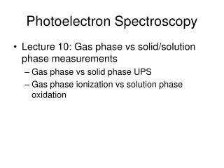
X-Ray Photoelectron Spectroscopy: Capabilities and Exploitation. Tim Morgan Mike Hawkridge. Outline. Photoemission Instrumentation Spectral Features Quantification Bonding States Depth Profiling Exploiting the XPS. Photoelectron Emission.

E N D
X-Ray Photoelectron Spectroscopy: Capabilities and Exploitation Tim Morgan Mike Hawkridge
Outline • Photoemission • Instrumentation • Spectral Features • Quantification • Bonding States • Depth Profiling • Exploiting the XPS
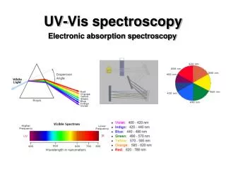
Photoelectron Emission • Photoelectric effect discovered by Einstein in 1905. Awarded Nobel Prize 1921. • Photoemission as an analytical tool by Kai Siegbahn. Awarded Nobel Prize 1981. KE = hν-BE-φs EVac • Spectrum is plot of electron count/ intensity versus Binding Energy or Kinetic Energy • Quantification of relativesurface composition (± X% (We’ll get to this!)) • Identification of element and chemical state (100ppm ultimate detection limit) • Valence Band Structure (UV best 10-45eV) φs EF photoelectron X-ray (hν) 2p BE 2s 1s
Photoelectron Emission KE = hν-BE-φs EVac EVac φan φs EF EF photoelectron Auger electron X-ray (hν) Analyzer 2p BE 2s KE = hν-BE-φs-(φan-φs) 1s Excited Ion Relaxed Ion KE = hν-BE-φan E(1s)-E(2s) available for either fluorescent x-ray or Auger emission. K-Shell emission
Typical Spectral Features • Sharp Photoelectron peaks • Broader Auger peaks with fine structure • Background from inelastically scattered photoelectrons
Photoemission Spectrometer SCA Lens Ar-ion C-60 Load Lock Main Chamber
Photoemission Spectrometer Quartz crystal monochromator V Energy Analyzer (SCA) Electron Gun Rowland Circle Al Kα x-rays (1486eV) Ion Gun 15-20kV electrons Lens E0 Electron Neutralizer MCD Al Anode 1eV electrons Photoelectrons Pass Energy Sample Resolution All in UHV for electron mfp and to minimize surface contamination.
Charge Neutralization X-ray beam Electron neutralizer +++ Conducting Sample
Charge Neutralization X-ray beam Electron neutralizer Ion Gun - - - - - - - - - - - - - - - - - - - +++ Insulating Sample
Quantification • Assume homogeneous composition throughout analysis volume. • Ii area under peak. • Ni number of ions, i. • σiphotoionization cross-section • λielectron attenuation length • K instrumental factors • First principles approach, or • Re-write equation for number of atoms: • Measure “pure” standards to determine sensitivity factors, Si • Can be done externally or internally Duke C B PNAS 2003;100:3858-3864
Sensitivity Factors in the VersaProbe • RSF Read from tables (inelastic MFP, photo-ionization xs buried in here). • Transmission function, T, in VersaProbe takes general form: • Measure Cu 3p (77eV), 2p3 (934eV) and LMM (568eV) peaks at various pass energies and fit to appropriate abscissa and ordinate. • Geometric correction: • β = asymmetry parameter for a specific atomic orbital of specific atom (tables) • θ = angle between x-ray and lens (= 54.7° = “magic” angle = VersaProbe angle)
Peak Area: Background Subtraction • Smooth the data if noisy • Subtract background due to inelastic scattering processes • Peak area (or height) is proportional to the atomic concentration cps si= bi+pi bi • (Iterated) Shirley background model most commonly used; Easy to apply and good enough for most practical purposes, if not physically accurate. • Source of error in concentration determination • Reliability decreases with less intense spectral peaks. KE Shirley Background Modeling
Error on Quantification • Sources: • Accuracy of relative sensitivity factors • Transmission function calibration • Accuracy of background determination • S/N ratio of data • Peak area included (must include all features associated with the subject peak, e.g. Plasmon loss peaks) • Generally accepted relative error of ±5% on atomic concentration • Larger for lower concentrations (lower reliability)
Depth Profiling - Sputtering C-60 Ar-ion • Ar- and C-60 ion guns can sputter surface material. • Cluster gun uses lower eV/atom and thus less surface damage (not energetic enough to sputter semiconductors) • Alternate sputtering for set time period and data collection (can also collect while sputtering). Tim’s Depth Profiling Data Polymer Profiling Data?
Depth Profiling – Angle Resolved XPS • Equation • Electrons escaping from deeper within the material are attenuated to a greater degree than those escaping from the surface. • Can use this fact and measure at varying escape angles to determine thin layer structure. • Highly accurate, but only for top ~10nm.