Download
1 / 29
300 likes | 545 Vues
Matt Rarick (X4-5225), matt.rarick@bmc.org. Yan Deng (X4-5225), ydeng@bu.edu. Gerald Denis (X4-1371), gdenis@bu.edu. Assays, Hardware and Software for Analysis and Advanced Cell Sorting. for users of the Flow Cytometry Core Facility at BUMC 21 October 2008.

E N D
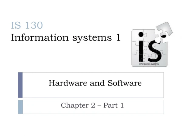
Matt Rarick (X4-5225), matt.rarick@bmc.org Yan Deng (X4-5225), ydeng@bu.edu Gerald Denis (X4-1371), gdenis@bu.edu Assays, Hardware and Software for Analysis and Advanced Cell Sorting for users of the Flow Cytometry Core Facility at BUMC 21 October 2008
Cell sorting applications GFP sorting Hoechst stain of “side populations” of stem cells Multicolor considerations From 10 to 15 colors and beyond Cell analysis applications Proliferation: Carboxyfluorescein succinimidyl ester (CFSE) Calcium flux: Indo dyes Viability: Live/dead discrimination Signaling: Phosphoflow Apoptosis: TUNEL stain
Cell sorting applications GFP sorting Hoechst stain of “side populations” of stem cells Multicolor considerations From 10 to 15 colors and beyond Cell analysis applications Proliferation: Carboxyfluorescein succinimidyl ester (CFSE) Calcium flux: Indo dyes Viability: Live/dead discrimination Signaling: Phosphoflow Apoptosis: TUNEL stain
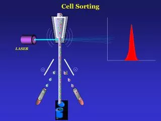
● low flow rate ● high flow rate ● narrow sample core ● wide sample core ● high resolution ● low resolution Which pressure/nozzle settings are optimal for my sort? sheath fluid ‘hydrodynamic focusing’ single file particles NOZZLE
Dependent variables Independent variables Yield/viability Nozzle size Time Fluid pressure Purity Cell size Which pressure/nozzle settings are optimal for my sort? We choose the nozzle size for your particular sort in consultation with you. Depends on the size and characteristics of the cells more than anything else. The nozzle size should be 4 - 5X that of the cells being sorted. lymphocytes: 70 mm nozzle larger cells: 100 mm nozzle Increased pressure and the smaller orifice of the 70 mm nozzle will expose your cells to a harsher shear, very briefly. If your cells are more delicate, we might choose the 100 mm nozzle regardless of their size. Bacteria can be sorted with an extremely narrow nozzle.
How long will my sort take? Typical low pressure sort: 100 mm nozzle 3000 cells/second = 10 million cells in one hour Typical high pressure sort: 70 mm nozzle 20,000 cells/second = 70 million cells in one hour We adjust the rate of cells/second based on the efficiency and quality of the sort, in relation to your needs and situation.
Sorting of GFP+ lentivirus-transduced cells Empty vector lentivirus GFP-lentivirus Visible microscopy Fluorescence microscopy Flow cytometry GFP FSC Courtesy of Wanda Blanton
Control Verapamil 0.25% Hoechst blue Hoechst red Identification of a Hoechst 33342-staining ‘side population’ from murine bone marrow 100% of the gated side population is also Sca1+ ; these are hematopoietic stem cells.
Kinetic identification of “side population” stem cells These stem cells can be isolated by MoFlo
Cell sorting applications GFP sorting Hoechst stain of “side populations” of stem cells Multicolor considerations From 10 to 15 colors and beyond: compensation issues Cell analysis applications Proliferation: Carboxyfluorescein succinimidyl ester (CFSE) Calcium flux: Indo dyes Viability: Live/dead discrimination Signaling: Phosphoflow Apoptosis: TUNEL stain
Problems in Emission Fluorescence Excitation Emission Spectral overlap
Filters resolve overlapping wavelengths of emitted light Longpass filter: transmits light of longer than or equal to a specific wavelength Shortpass filter: transmits light of shorter than or equal to a specific wavelength Bandpass filter: transmits light only within a narrow range of wavelengths Optical solutions to spectral overlap: Filters
EMISSION two bandpass filters
Uncompensated Optimal Electronic solutions to spectral overlap: Compensation To correct for emission spillover of FITC signal (normally detected in the FL1 channel) into the FL2 channel (which detects PE), it is necessary to use filters or electronic compensation or both.
Compensation Multicolor immunophenotyping Before After
Filter configurations permit optimization of multicolor stains
Qdot nanocrystals offer an option to increase the number of usable emission wavelengths Qdot 655 Qdot 525 Qdot 565 Qdot 585 Qdot 605 Qdot 705
100 100 80 80 60 60 % of Max % of Max 40 40 20 20 0 0 0 1 2 3 4 0 1 2 3 4 10 10 10 10 10 10 10 10 10 10 100 100 80 80 60 60 % of Max % of Max 4 10 40 40 3 20 20 10 0 0 2 0 1 2 3 4 0 1 2 3 4 10 10 10 10 10 10 10 10 10 10 10 1 10 0 10 0 200 400 600 800 1000 Multicolor, Auto Compensation with Flow Jo isotype isotype gate 1: lymphocytes gate 2: B cells gate 6: T cells? FITC Pacific Blue isotype isotype etc gates 3 – 5 up to 14 colors PE PE-Cy7
How does the LSR II work? Octagon (Blue 488 nm laser) SSC 585/42 488 PE 550LP 635LP PerCP-Cy5.5 695/40 780/60 PE-Cy7 685LP incident light 505LP FITC 530/30
Trigon (Violet 405 nm laser) 505LP 610/20 Pacific Blue empty 450/50 Qdot 605 Trigon (UV 325 nm laser) 450LP 735LP 780/60 450/40 APC Live/Dead empty empty 660/20 APC-Cy7 Alexa 350 405/20 Trigon (Red 633 nm laser) 3 Trigon configurations
FITC Compensation Matrix FITC FITC
Cell sorting applications GFP sorting Hoechst stain of “side populations” of stem cells Multicolor considerations From 10 to 15 colors and beyond Cell analysis applications Proliferation: Carboxyfluorescein succinimidyl ester (CFSE) Calcium flux: Indo dyes Viability: Live/dead discrimination Signaling: Phosphoflow Apoptosis: TUNEL / caspase-3 / Annexin V / PI stains
Assay of proliferation by dilution of CFSE label 3 2 0 4 cycles of cell division 1 0 curve fitting algorithms
Live and dead cells distinguished by flow cytometry blue violet red far red aqua green near IR
“Phosphoflow” techniques allow you to measure kinase cascades and signal transduction Courtesy John Meyers Lerner Lab EBRC 4 johnm@bu.edu
Assays of apoptosis TUNEL vs. caspase-3 TUNEL Annexin V vs. propidium iodide
Matt Rarick (X4-5225), matt.rarick@bmc.org Yan Deng (X4-5225), ydeng@bu.edu Gerald Denis (X4-1371), gdenis@bu.edu