Download
1 / 65
660 likes | 868 Vues
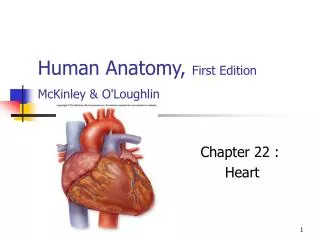
Human Anatomy, First Edition McKinley & O'Loughlin. Chapter 22 : Heart. Functions of the Heart. Center of the cardiovascular system, the heart. Connects to blood vessels that transport blood between the heart and other body tissues. arteries carry blood away from the heart

E N D
Human Anatomy, First EditionMcKinley & O'Loughlin Chapter 22 : Heart
Functions of the Heart • Center of the cardiovascular system, the heart. • Connects to blood vessels that transport blood between the heart and other body tissues. • arteries carry blood away from the heart • veins carry blood back to the heart • Arteries carry blood high in oxygen. • (except for the pulmonary arteries) • Veins carry blood low in oxygen. • (except for the pulmonary veins) • Arteries and veins entering and leaving the heart are called the great vessels.
Characteristics and Functions of the Heart • Ensures the unidirectional flow of blood through both the heart and the blood vessels. • Backflow of blood is prevented by valves within the heart. • Acts like two independent, side-by-side pumps that work independently but at the same rate. (double circuit) • one directs blood to the lungs for gas exchange • the other directs blood to body tissues for nutrient delivery
Characteristics and Functions of the Heart • Develops blood pressure through alternate cycles of heart wall contraction and relaxation. • Minimum blood pressure is essential to push blood through blood vessels to the body tissues for nutrient and waste exchange.
Pulmonary and Systemic Circuits • The pulmonary circuit consists of the chambers on the right side of the heart (right atrium and ventricle) as well as the pulmonary arteries and veins. • conveys blood to the lungs via pulmonary arteries • to reduce carbon dioxide and replenish oxygen levels in the blood • Blood returns to the heart in pulmonary veins
Pulmonary and Systemic Circuits • Blood returns to the left side of the heart, where it then enters the systemic circuit. • The systemic circuit consists of the chambers on the left side of the heart (left atrium and ventricle), along with all the other named blood vessels. • carries blood to all the peripheral organs and tissuesof the body
Pulmonary and Systemic Circuits • Oxygenated blood from the left side of the heart is pumped into the aorta • the largest systemic artery in the body • then into smaller systemic arteries. • Gas exchange in tissues occurs from capillaries. • Systemic veins then carry deoxygenated blood (high in carbon dioxide) and waste products. • Most veins merge and drain into the superior and inferior venae cavae • drain blood into the right atrium. • There, the blood enters the pulmonary circuit, and the cycle repeats .
Anatomy of the Heart • Relatively small, conical organ approximately the size of a person’s clenched fist. • it weighs about 250 to 350 grams • Located left of the body midline posterior to the sternum in the middle mediastinum. • Rotated such that its right side or border (right atrium and ventricle) is located more anteriorly, while its left side or border (left atrium and ventricle) is located more posteriorly.
Anatomy of the Heart • The posterosuperior surface of the heart, formed primarily by the left atrium, is called the base. • The pulmonary veins that enter the left atrium border this base. • The inferior, conical end is called the apex. • It projects slightly anteroinferiorly toward the left side of the body.
Pericardium • Fibrous, serous sac • Contains the heart • In the mediastinum • Held in place by connective tissue • The external wall of the great vessels’ superior to the heart • diaphragm inferior. • Restricts heart movements • Prevents the heart from overfilling with blood.
Pericardium • Outer portion • tough, dense connective tissue • called the fibrous pericardium. • attached to both the sternum and the diaphragm • Inner portion • thin, double-layered serous membrane • called the serous pericardium. • parietal layer • visceral layer
Heart Wall Structure • Three distinctive layers: • external epicardium • middle myocardium • internal endocardium • Epicardium • outermost heart layer • also known as the visceral layer of serous pericardium. • Simple squamous epithelium underlined by fat • As we age, more fat is deposited in the epicardium • this layer becomes thicker and more fatty.
Heart Wall Structure • Myocardium • middle layer of the heart wall • composed chiefly of cardiac muscle tissue. • thickest of the three heart wall layers. • lies deep to the epicardium and superficial to the endocardium • Endocardium • covers internal surface of the heart and the external surfaces of the heart valves • thin endothelium • areolar CT under the endothelium
Cardiac Muscle Tissue • Fiber Characteristics • short, branched fibers • one or two central nuclei • numerous mitochondria for ATP supply. • striated, with extensive capillary networks • Fiber arrangement • in spiral bundles • wrapped around and between the heart chambers.
Cardiac Muscle Tissue • Fibers contract as a single unit • Intercalated discs • Specialized cell–cell contacts • Contain gap junctions • contain desmosomes • Muscle impulses are distributed immediately and simultaneously throughout all fibers either of the atria or of the ventricles.
External Anatomy of the Heart • Chambers: • four hollow chambers: • two smaller atria • two larger ventricles. • Atria • thin-walled, located superiorly. • anterior part of each atrium is a wrinkled, flaplike extension called an auricle • Atriareceive blood through both circulatory circuits. • right atrium receives blood from the systemic circuit • left atrium receives blood from the pulmonary circuit
External Anatomy of the Heart • Blood that enters an atrium is passed to the ventricle on the same side of the heart. • Ventricles • the inferior chambers. • Two large arteries, the pulmonary trunk and the aorta exit the heart at the basal surface. • The pulmonary trunk carries blood from the right ventricle into the pulmonary circuit. • The aorta conducts blood from the left ventricle into the systemic circuit
External Anatomy of the Heart • Atria are separated from the ventricles externally by coronary sulcus (or atrioventricular sulcus) • extends around the circumference of the heart. • On both the anterior and posterior surfaces of the heart, the anterior interventricular sulcus and the posterior interventricular sulcus are located between the left and right ventricles. • These sulci extend inferiorly from the coronary sulcus toward the heart apex.
Functions of the Fibrous Skeleton of the Heart • Located between the atria and the ventricles • Formed from dense irregular connective tissue. • separates the atria and ventricles • anchors heart valves by forming supportive rings at their attachment points • provides electrical insulation between atria and ventricles • ensures that muscle impulses are not spread randomly throughout the heart • prevents all of the heart chambers from beating at the same time • Provides a rigid framework for the attachment of cardiac muscle tissue.
Internal Anatomy of the Heart • There are four heart chambers: • right atrium • right ventricle • left atrium • left ventricle • Each plays a role in the continuous process of blood circulation. • Valves permit the passage of blood in one direction and prevent its backflow.
Right Atrium • Receives venous blood • from the systemic circuit • from the heart muscle itself. • Three major vessels empty into the right atrium: • superior vena cava (SVC) • drains blood from the head, upper limbs, and superior regions of the trunk • inferior vena cava (IVC) • drains blood from the lower limbs and trunk • coronary sinus drains blood from the heart wall • The interatrial septum forms a wall between the right and left atria.
Right Atrioventricular (AV) Valve • Separates the right atrium from the right ventricle. • Also called the tricuspid valve. • has three triangular flaps • Venous blood flows from the right atrium, through the valve into the right ventricle. • Is forced closed when the right ventricle begins to contract • preventing blood backflow into the right atrium
Right Ventricle • Receives deoxygenated venous blood from the right atrium. • An interventricular septum forms a wall between the right and left ventricles. • Papillary muscles • on the internal wall surface • cone-shaped, muscular projections • anchor chordae tendineae • attach to the cusp of the right AV valve and prevent everting and flipping into the atrium when contracting
Pulmonary Trunk • At its superior end it narrows into a smooth-walled, conical region called the conus arteriosus. • The pulmonary semilunar valve marks the end of the right ventricle and the entrance into the pulmonary trunk. • Pulmonary trunk divides shortly into right and left pulmonary arteries. • carry deoxygenated blood to the lungs
Semilunar Valves • Located within the walls of both ventricles • immediately before the connection of the ventricle to the pulmonary trunk and aorta. • Composed of three thin, pocketlike semilunar cusps. • As blood is pumped into the arterial trunks, it pushes against the cusps • forcieg the valves open. • When ventricular contraction ceases • blood is prevented from flowing back into the ventricles. • causes the cusps to “inflate” and meet at the artery center, effectively blocking blood backflow
Left Atrium • Once gas exchange occurs in the lungs, the oxygenated blood travels through the pulmonary veins to the left atrium. • Smooth posterior wall of the left atrium contains openings for approximately four pulmonary veins. • two left pulmonary veins • two right pulmonary veins • Has pectinate muscles along its anterior wall as well as an auricle.
Left Atrioventricular (AV) Valve • Separates the left atrium from the left ventricle. • Also called the bicuspid valve or the mitral valve. • Left AV valve has chordae tendineae similar to those of the right AV valve. • Oxygenated blood flows from the left atrium into the left ventricle. • Is forced closed when the left ventricle begins to contract • prevents blood backflow into the left atrium
Left Ventricle • Largest of the four heart chambers. • Wall is typically three times thicker than the right ventricular wall. • Requires thick walls in order to generate enough pressure to force the oxygenated blood from the lungs into the aorta and then through the entire systemic circuit. • right ventricle only has to pump blood to the nearby lungs
Left Ventricle • Trabeculae carneae in the left ventricle are more prominent. • Two large papillary muscles attach to the chordae tendineae that help support the left AV valve. • At the superior end of the ventricular cavity, the aortic semilunar valve marks the end of the left ventricle and the entrance into the aorta.
Cardiac Cycle • The inclusive period of time from the start of one heartbeat to the initiation of the next. • All chambers within the heart experience alternate periods of contraction and relaxation. • Contraction of a heart chamber is called systole. • forces blood into another chamber (from atrium to ventricle) • forces blood into a blood vessel (from a ventricle into the attached large artery) • Relaxation phase of a heart chamber is termed diastole. • myocardium of each chamber relaxes between contraction phases • and the chamber fills with blood
Conduction System of the Heart • Exhibits autorhythmicity • the heart itself (not external nerves) is responsible for initiating the heartbeat. • Certain cardiac muscle fibers are specialized to conductmuscle impulses to the contractile muscle cells of the myocardium. • Specialized cells are part of the heart’s conduction system.