Smooth Muscle
120 likes | 257 Vues
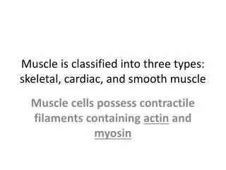
Smooth Muscle. Prof. K. Sivapalan. Smooth Muscle. Found in the internal organs Usually 1 to 5 µ in diameter and only 20 to 500 µ in length Myosin and actin filaments cause contraction but the arrangement is different. Smooth M uscle Types.

Smooth Muscle
E N D
Presentation Transcript
Smooth Muscle Prof. K. Sivapalan
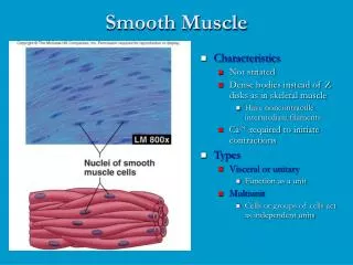
Smooth Muscle • Found in the internal organs • Usually 1 to 5 µ in diameter and only 20 to 500 µ in length • Myosin and actin filaments cause contraction but the arrangement is different
Smooth Muscle Types • The smooth muscle of each organ is distinctive in several ways: • (1) physical dimensions, • (2) organization into bundles or sheets, • (3) response to different types of stimuli, • (4) characteristics of innervation, and • (5) function.
Major Types • Multi Unit Smooth Muscle: • Discrete fibers • Each operate independently • Main control is nervous • Examples: ciliarymuscle of the eye, the iris muscle of the eye, and the piloerector muscles
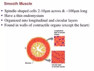
Major Types • Unitary Smooth Muscle [Visceral Smooth Muscle]: • All act as single unit • Arranged in bundles or sheets • Fibers are attached so that the force of contraction is transmitted from to the next • Syncytial- communicate through gap junctions. • examples: the gut, bile ducts, ureters, uterus, and blood vessels
Innervation • Smooth muscles are innervated by autonomic fibers. • The nerves end in varicosities which release the transmitter: • Sympathetic- noradrenaline [norepinephrine] • Parasympathetic- acetyl choline
Arrangement of Filaments • Actin filaments are attached to dense bodies • Dense bodies are distributed in the cytoplasm and the cell membrane. • Dense bodies in the membrane of adjuscent cells are bound to transmit the force. • Myosin filaments are interspersed among actin • 80 % shortening is possible
Electrical Property • RMP is -50 to -60 mV • When stimulated spike potentials occur, in some with plateau [uterus]. • In some unitary muscles, slow wave rhythm is seen. • When this spontaneous change reaches the threshold level, spike potential occurs. [pace makers in GIT]
Action Potentials • Duration of spike potentials- 10-50 ms • Where there is a plateau, it may last up to 1000 ms • The depolarization is mostly due to voltage gated calcium channels [which are slow to open] than sodium channels.
Stimulation • Stimulation may or may not result in action potential. The factors react with receptors to cause contraction or relaxation. • Local chemical factors: • Hypoxia, hypercapnoea, acidity, lactic acid, adenosine, potassium ions, NO and temperature • Hormones: • Epinephrine, norepinephrine, acetyl choline, angiotensin, endothelin, oxytocin, serotonin, histamine • The response depends on the receptors. • Stretch
Excitation-Contraction Coupling • Calcium binds to calmodulin [no troponin] • It activates myosin kinase • Regulatory chain of myosin head is phosphorilated • Binding with actin repeatedly • When calcium is removed, myosin phosphatase is activated. • It splits the phosphate from myosin head and contraction ceases
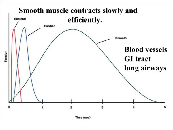
Mechanical Property • The oncet of contraction is slow and the contraction time is prolonged. • Energy required is also very much less • The force of contraction per unit surface area is much more- 4-6 kg/cm2 [3-4 in skeletal muscles]