Download
1 / 36
380 likes | 560 Vues
Abnormal EEG brain in neurological disease. แพทย์หญิง กาญจนา พิทักษ์วัฒนานนท์ อายุรแพทย์ผู้เชี่ยวชาญระบบประสาท แพทย์ประจำศูนย์สมอง โรงพยาบาลสมิติเวชศรีราชา. 10 – 20 system. Records the difference between two Active scalp electrodes. Bipolar montage. Active scalp electrode

E N D
Abnormal EEG brain in neurological disease แพทย์หญิง กาญจนา พิทักษ์วัฒนานนท์ อายุรแพทย์ผู้เชี่ยวชาญระบบประสาท แพทย์ประจำศูนย์สมอง โรงพยาบาลสมิติเวชศรีราชา
Records the difference between two Active scalp electrodes Bipolar montage
Active scalp electrode • Inactive scalp / ear electrode : A1 , A2 Referential montage
If the voltage input from electrode 2 is relatively “ positive ” compared to electrode 1 , then the pen will deflect “ up ” • If electrode 2 is relatively “ negative ” compared to electrode 1 , then the pen will deflect “ down ” • If electrode 1 equal to electrode 2 , then the pen stay “ baseline ” Polarity
Identify background rhythms ( frequency ) • Alpha 8 -13 Hz • Beta 13 – 40 Hz • Theta 4 – 8 Hz • Delta < 4 Hz • Mu 7 – 11 Hz • Sleep stage ( awake , NREM , REM ) • Identify any abnormal patterns • Slow patterns • Diffusely , Focally • Symmetry , Asymmetry • Paroxysms • Spike , spike and wave complex , sharp wave • Triphasic waves • PLEDs ( Periodic lateralizing epileptiform discharges ) • Localize those abnormal patterns • Maximum amplitude ( Referential montage ) • Phase reversal / Reciprocal changes ( Bipolar montage ) EEG interpretation
Common • Sleep deprivation , • Alcohol withdrawal , • Television flicker , • Systemic infection , Head trauma , • Recreational drugs , antiepileptic drug , • Menstruation • Occasional • Dehydration , • Barbiturate / Benzodiazepine withdrawal , • Hyperventilation, • Flashing lights , • Diet and missed meals , • Specific reflex trigger , • Stress , Intense exercise Factor lowering seizure threshold
Rountine electroencephalograms • Hyperventilation • Photic stimulation • Sleep-deprivation • Prolonged ambulatory recording • Video – EEG telemetry • Magnetoencephalography : Research Activation tecniques ( EEG )
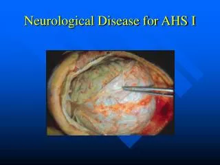
Herpes simple encephalitis • PLEDs ( Periodic lateralizing epileptiform discharges )
Absence seizure 3 Hz spike and wave complexs
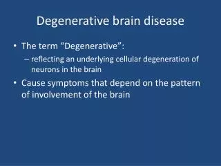
CJD – Mad cow disease Diffused slow pattern : encephalopathy
Prion encephalitis • Triphasicwaves
Severe dementia • Triphasicwaves : degenerative disease