Abdominal and Thoracic Effusions
Abdominal and Thoracic Effusions. Clinical Pathology. Abdominal/thoracic fluids. Abdominal and thoracic organs are bathed in and lubricated by a small amount of fluid Fluid increases when the amount entering the cavity is more than is removed fro it

Abdominal and Thoracic Effusions
E N D
Presentation Transcript
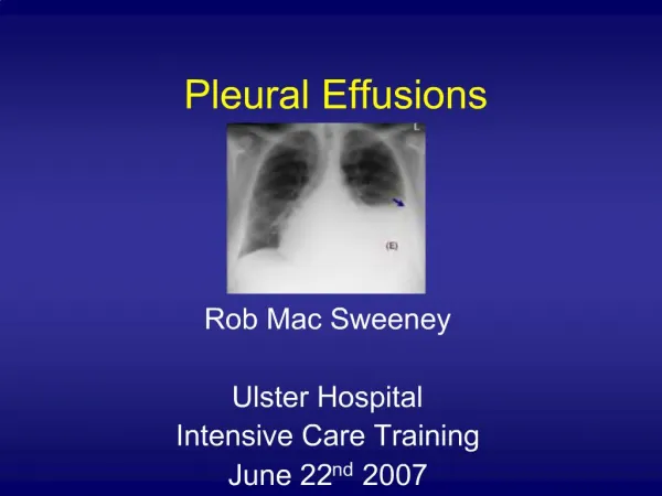
Abdominal and Thoracic Effusions Clinical Pathology
Abdominal/thoracic fluids • Abdominal and thoracic organs are bathed in and lubricated by a small amount of fluid • Fluid increases when the amount entering the cavity is more than is removed fro it • An increased amount of fluid in the abdominal/thoracic cavity is not a disease in itself, but rather an indication of a pathologic process in the fluid production and/or removal system.
Abdominal/Thoracic Effusion • Fluid analysis and cytology is quick, easy and inexpensive, and relatively safe. • May obtain useful information for diagnosis, prognosis, and treatment.
Abdominocentesis Collection • Aseptic prep of the skin • Usually done with animal standing • Use a sterile needle or cannula • Tap the ventral midline of the abdomen, 1-2 cm caudal to the umbilicus • Collect fluid and place into an EDTA and red top tube
Indications of a peritoneal tap • Ascites: due to cardiac or liver disease or neoplasia, etc. • Peritonitis: Ruptured bowel, ruptured bladder. • In horses-colic
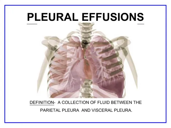
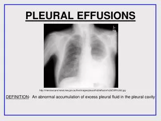
Thoracentesis • Pleural effusion may be bilateral or unilateral. • Radiographs help determine the extent and location. • Usually place in sternal recumbency. • Aseptic prep. • Sterile needle, catheter inserted next to cranial surface of the rib to prevent risk of penetrating the vessel on the caudal border of the rib.
Indications for Thoracentesis • Hemorrhage • Inflammation (FIP) • Neoplasia • Ruptured thoracic lymphatic duct (chylothorax) Place in EDTA and Red top tube
Characteristics of Effusions • Transparency/turbidity • Color • Protein concentration • Specific gravity • Cells: counts, types, and morphology • Fluid should be odorless- if an abdominal tap yields a malodorous fluid- may indicate a ruptured bowel
Color/turbidity of Effusions • Influenced by protein concentrations and cell numbers. • Normal peritoneal/pleural fluid is colorless and transparent to slightly turbid. • FIP may cause an amber, turbid, thick effusion (straw-yellow color)
Total Protein and Specific Gravity • Centrifuge sample at low speed (1500 rpm for 5 min) • TP can be measured on refractometer • Normal is <2.5 g/dl • Specific gravity is measured on refractometer as well • Normal is <1.018
Slide preparation • Centrifuge fluid at low speed • Pour off supernatant, leave about 0.5 ml at the bottom of tube • Resuspend by gentle agitation • Place a drop on slide • Routine blood smear type technique • Squash prep • Air dry • Stain with diff quick
Cell counts on Effusions • Total nucleated cell counts • Unopette procedure • Automated • Normal peritoneal/pleural fluid has <10,000 nucleated cells/ul • Estimated cell counts can be made on a blood smear
Types of cells found in effusions • Neutrophils • Mesothelial/macrophage type cells • Lymphocytes • Eosinophils • Mast cells • Neoplastic cells
Classifications of Effusions • Transudates • Modified transudates • Exudates
Transudates • Clear, colorless effusion • <2.5 g/dl protein (low protein) • Low total nucleated cell count • Non-degenerative neutrophils and mesothelial cells • Specific gravity < 1.013
Causes of Transudates • Hypoalbunemia: due to renal glomerular disease, hepatic insufficiency, and protein-losing enteropathy. • Ruptured bladder • Rarely from blockage of lymph from lymphatic vessel in the intestines
Modified Transudates • Vary in color- amber to white to red • Frequently slightly turbid to turbid • High protein concentration (2.5-7.5 g/dl) • Moderate cellularity: 1000-7000 cells/ul • Occur as a result of fluid leakage from lymphatics carrying high protein lymph or blood vessels.
Modified transudate causes • Lease specific, variety of disorders • Cardiovascular disease (right sided heart failure) • Neoplastic disease • FIP • Chylothorax • Hemorrhage • Hepatic disease- hypoalbunemia and hypertension
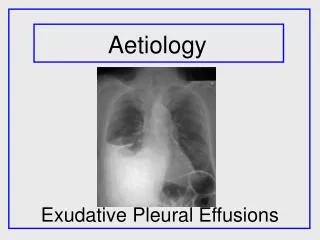
Exudates (infections) • Color varies- amber to white to red • Turbid to cloudy • High protein > 3.0 g/dl • High total nucleated cell count (>7000 cell/ul) • Neutrophils are the predominant cell type.
Exudates continued • Septic exudates: Degenerative neutrophils and intracellular/extracellular bacteria present • Ex: GI perforation, systemic sepsis, pneumonia • Non-septic exudates: non-degenerative neutrophils, small lymphocytes, and/or neoplastic cells. • Ex: Pancreatitis, neoplasia, uroperitoneum
Chylous Effusions • Contains chylomicron-rich fluid that is present in lymphatics that drain the intestinal tract and pass through the thoracic duct. • Chylomicrons are triglyceride-rich lipoproteins absorbed from the intestines after the ingestion of food containing lipids. • Chyle normally drains from the thoracic duct into the venous system. • Effusion forms when there is an obstruction of the lymphatic flow resulting in increases pressure within the lymphatics and dilation of the thoracic duct. • Other causes included: cardiovascular disease or trauma • Lymphocytes are predominant type