Download
1 / 1
30 likes | 166 Vues
University of Wisconsin-Madison Materials Research Science & Engineering Center on Nanostructured Interfaces. UW MRSEC DMR-0520527 Juan J. de Pablo, PI. Early Detection of Amyloid Fibrils by Liquid Crystals. Aslin Izmitli, Nicholas Abbott, and Juan de Pablo .

E N D
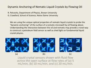
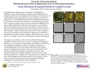
University of Wisconsin-Madison Materials Research Science & Engineering Center on Nanostructured Interfaces UW MRSEC DMR-0520527 Juan J. de Pablo, PI Early Detection of Amyloid Fibrils by Liquid Crystals Aslin Izmitli, Nicholas Abbott, and Juan de Pablo Neurodegenerative diseases such as Alzheimer’s, Huntington’s or Parkinson’s disease afflict millions of patients in the United States and around the world. One of the hallmarks of such diseases is the formation of amyloid fibrils; protein aggregates that appear as plaques or deposits in damaged tissue. An important challenge in devising therapeutic strategies for such diseases has been early detection of such fibrils and the disease itself. A group of scientists at the University of Wisconsin MRSEC led by Juan de Pablo and Nicholas Abbott has demonstrated a novel amyloid-fibril detection platform based on liquid crystalline materials. Amyloid fibrils are rich in beta-sheet structures; when a liquid crystal is exposed to such structures, it adopts distinct patterns and textures that are unique to beta sheets. The liquid crystal can detect the appearance of such fibrils much earlier than other available techniques, such as fluorescence, and it can do so by relying solely on fast and inexpensive optical measurements. The figure on the right (a,b,c) shows polarized light micrographs of amyloid-beta fibrils as they grow (as a function of time) on a lipid layer, which serves as a model for a cell membrane. Such fibrils are believed to be responsible for Alzheimer’s disease. The figure also shows the corresponding images (g-i) after staining with a fluorescent dye that reports the formation of beta sheets. One can see that the liquid crystal provides a faithful representation of the image generated by more demanding fluorescence labeling techniques, and it can do so at much smaller concentrations or length scales, when fluorescence simply does not work. The technique has been demonstrated on a wide variety of other disease-causing, fibril forming proteins, including polyglutamine (Huntington’s disease) and amylin (type II diabetes). Figure Caption: Optical response of the lipid-laden interface of LC to Aβ adsorption. (a-c) are polarized light micrographs, (d-f) are phase contrast images and (g-i) are epifluorescence micrographs of texas red labeled Aβ. Panel (j) shows the epifluorescence micrograph of ThT fluorescence at the end of the experiment. The images are taken at 5 minutes (a, d, g), 30 minutes (b, e, h) and 1 hour and 30 minutes (c, f, i, j) after the addition of the peptide into the aqueous phase. The scale bar is 100μm. Aslin Izmitli, Nicholas Abbott, and Juan de Pablo, submitted for publication, 2010.