Dr W Kolbinger, Visual System (2009)
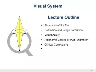
Visual System. Lecture Outline. Structures of the Eye Refraction and Image Formation Visual Acuity Autonomic Control of Pupil Diameter Clinical Correlations . 1. 1. Dr W Kolbinger, Visual System (2009). Anatomic Considerations. The Ocular Fundus. Optic disc. Fovea. Macula.

Dr W Kolbinger, Visual System (2009)
E N D
Presentation Transcript
Visual System Lecture Outline • Structures of the Eye • Refraction and Image Formation • Visual Acuity • Autonomic Control of Pupil Diameter • Clinical Correlations 1 1 Dr W Kolbinger, Visual System (2009)
The Ocular Fundus Optic disc Fovea Macula The optic disc region itself only contains axons of retinal ganglion cells, the output elements of the retina, but it lacks photoreceptors. As a consequence, the optic disc is responsible for the blind spot, a region inside the boundaries of the visual field, where we don’t receive visual information. 3
Optics of the Eye Convex Lens Focuses Light Rays Concave Lens Diverges Light Rays Measurement of the Refractive Power of a Lens—“Diopter” The refractive power in diopters of a convex lens is equal to 1 meter divided by its focal length.
Optics of the Eye Cornea refractive power: 42 D Flat lens refractive power: 13 D Rounded lens refractive power: 26 D Plasticity: 13 D 5
Accommodation Flat lens refractive power: 13 D • Far Vision Ciliary muscle relaxed Suspensory ligaments tightened Focus on the Retina Accommodation Adjusts the Refractive Power of the Eye 6
Accommodation Rounded lens refractive power: 26 D • Near Vision Ciliary muscle constricted Suspensory ligaments floppy Focus on the Retina 7
Presbyopia Flat lens • Near Vision The variability of the refractive power of the lens between far vision (13 D) and near vision (26 D) is called refractive plasticity. Unfortunately, the lens looses its elasticity during aging, thereby reducing the ability to focus on near objects, a condition called presbyopia. Blurred picture on the Retina 8
Emmetropia (Normal Vision). Cataracts “Cataracts” are an especially common eye abnormality that occurs mainly in older people. A cataract is a cloudy or opaque area or areas in the lens
Visual acuity is the ability to distinguish between two nearby points. Visual acuity is high when the two-point discrimination threshold is low (high spatial resolution). What is Visual Acuity? • Two point discrimination of the visual system • Normal: an angle of 5 minutes of a degree • highly dependent on the densities of retinal photoreceptors visual acuity also depends on a proper function of the optical apparatus of the eye, including accommodation. When the optical apparatus fails to produce a focused (sharp) picture on the retina, the objects in the visual field appear “blurry”. 5 ‘ Far vision Near vision 10
Neurological Examination of Visual Acuity 20 800 Distance equivalents 20 20 • Eye charts and near cards Patient Normal 11
RETINA- Receptor and Neural Function of the Retina • Photoreceptors and Phototransduction • Color Vision • Processing of Visual Information • Clinical Correlations 13 13 Dr W Kolbinger, The Retina (2009)
Layers of the Retina Photoreceptor layer
Photoreceptors Outer segment • -Visual pigment Inner segment • Synapses with bipolar • and horizontal cells rhodopsin Disks Nucleus Synaptic ending Rods are highly sensitive to light and enable us to see under low intensity light conditions (at night). Cones(3) are less sensitive to light. enable us to see colors. glutamate Rod Cone 15
Convergence is high in the rod • system. It is low in the cone system. As a consequence, spatial resolution (visual acuity) is better in bright light, when the cone system is active. • Rods and cones are not evenly distributed over the whole retina and the fovea only contains cones, but no rods. As a consequence, there is no central vision under dim light conditions • Night Blindness
The Visual Pigment Rhodopsinis the visual pigment of rods. It consists of two components: · Opsin, a protein which is synthesized in the photoreceptor (cones have different types of opsins). · Retinal, a chromophore, is the light absorbing compound or the visual pigment. It is derived from Vitamin A and is the chromophore of the visual pigment in rods and cones. Vitamin A is synthesized from beta-carotene contained in our food.
Photoreception -The Dark Current GDP , cGMP Na+ G protein (Transducin) cGMP-gated channel • Visual pigment-Rhodopsin cGMPphosphodiesterase 18 Dr W Kolbinger, The Retina (2009)
Phototransduction GTP cGMP Na+ 5’GMP cGMPphosphodiesterase Light 19 Dr W Kolbinger, The Retina (2009)
Color Vision Is Based on Comparison of Activity of Three Cone Types Visible Light Is Part of the Electromagnetic Spectrum The visible part of the spectrum is characterized by wavelengths ranging from 400 to 700 nm (nanometers). 20
Monochromatic Light: Colors of a Rainbow 21 Dr W Kolbinger, The Retina (2009)
Three Types of Cones Have Different Spectral Sensitivities The human retina contains three types of cones: · S (short wavelength sensitive) cones, also called “blue” cones, with a maximum sensitivity at 430 nm · M (medium wavelength sensitive) cones, also called “green” cones, with a maximum sensitivity of 530 nm · L (long wavelength sensitive) cones, also called “red” cones, with a maximum sensitivity of 560 nm.
Stimulation of Cones by Monochromatic Light Log relative sensitivity 450 nm monochromatic light 400 500 600 700 Wavelength (nm) 23
Stimulation of Cones by Monochromatic Light 600 nm monochromatic light Log relative sensitivity 400 500 600 700 Wavelength (nm) 24
Red-Green Color Blindness Normal Protanopia Deuteranopia Individuals affected by red-green color blindness can no longer distinguish certain red colors from certain green colors. 25
pseudo-isochromatic color plates like the one on the right are presented to the patient. The numbers embedded in the pattern of colored dots can be distinguished by individuals with a fully intact color vision. Dichromats, who are weak in red-green discrimination, have difficulties in identifying the numbers on all plates
Visual Pathways Lecture Outline • The Visual Field • Passage of Light • Neuronal Pathways • The Primary Visual Cortex • Parallel Pathways for Depth, Motion, Form and Color • Clinical Correlations 27
The Visual Field Total amount of space we can see with this eye, when the eye is fixed straight ahead, pointing towards the center of the visual field (point of fixation). The extension of the visual field is measured in degrees of maximum deviation from this straight line in all directions. For right eye- - left visual hemifield/nasal - right visual hemifield/temporal The vertical axis together with the horizontal axis divide the visual field into Four quadrants:- superior left, superior right, inferior left and inferior right quadrant of the visual field.
Projections of the Visual Field on the Retina The four quadrants of the visual field are projected onto the retina. The superior half of the visual field is projected to the inferior half of the retina, and vice versa. The left half of the visual field is projected on the right half of the retina, and vice versa. The nasal visual hemifieldof the right eye temporal hemiretinaof the right eye. temporal visual hemifield of the right eye nasal hemiretina of the right eye visual field defects e.g “bitemporalhemianopia`` 29
Chief Complaint: Headache and Nausea History: A 76 year old retired college dean presents with recurrent headaches over the past four years. Her headaches have significantly increased in duration and intensity over the past few months. She also reports episodes of nausea and vomiting. Her husband adds that she has recently developed difficulties with comprehending spoken language. The patient experiences uncoordinated movements of the right hand. She stopped drinking and smoking cigarettes during her first pregnancy, when she was 27. General Examination: 76 year old female in no acute distress. No significant cardiac, respiratory, or abdominal abnormalities. Vital signs unremarkable. Neurological Examination: The patient can speak fluently, but the content is frequently incomprehensible to the listener. Her ability to read and write is markedly reduced (dyslexia and dysgraphia respectively.) The patient can not identify a hairbrush, but was able to demonstrate its use. Numbness was detected in the right lower face and right hand. In addition, the examination of the right hand revealed impaired 2-pt discrimination, joint position, and fine touch. Motor examination and reflex testing of the lower extremities were unremarkable. Marked weakness of right arm flexion and extension was present, as well as elevated right biceps and brachioradialis reflexes.
Neuronal Pathways, From the Retina Onwards relay station between the retina and the primary visual cortex. circadian clock afferent limb of the pupillary light reflex contribute to eye movements
The Primary Visual Cortex V1 Occipital pole Area 17 34
The Primary Visual Cortex V1 Calcarine sulcus Parieto-occipital sulcus Area 17 35
Neuronal Pathways, From the Retina Onwards • Fibers originating in nasal hemiretina cross over at the optic chiasm, fibers • originating in the temporal hemiretina don’t. • LGN fibers carrying sensory information of the superior half of the visual field follow the temporal radiation pathway, fibers originating in the inferior half of the visual field follow the parietal radiation pathway. Example1- the axons originating in the right LGN which are carrying sensory information from the superior left quadrant of the visual field, use the temporal radiation and synapse in the inferior portion (below the calcarine sulcus) of the primary visual cortex (V1). Example 2 - ?
Retinotopic Organization of V1 Visual field left eye Visual field right eye Left hemisphere Right hemisphere 37
Neurological Examination of the Visual Fields The visual pathways are commonly tested in neurological examinations and they have high localizing value. Loss of vision is clinically tested in each quadrant of the visual field in a “confrontation visual field test”. In this test, each eye is tested separately by having the patient look straight at the examiners eye, while standing in double arms length distance. While the examiner occludes his left eye with one hand, the patient occludes his right eye (and vice versa). Then the examiner moves his other hand, with one (or more) of his fingers stretched out, gradually from the periphery to the center of the visual field, to determine where it is first seen. Assuming the examiner has normal vision, the patient should see the appearance of the hand at the same time as the examiner. He should also be able to tell the number of fingers stretched.
Sparing of the Macula When the macula is not included, Macular sparing is often associated with vascular lesions involving the posterior cerebral artery or its branches,blood supply of the occipital pole of the cerebbral cortex (the area representing macular vision) may stay intact, due to sufficient blood flow from the middle cerebral artery.
Eye Movements Lecture Outline • Types of Eye Movements • Extraocular Muscles, their Innervation and Control • Saccadic Eye Movements • Clinical Correlations 40
Types of Eye Movements • Conjugate Eye Movements • Saccadic eye movements (and gaze) • Vestibulo-ocular reflex • Optokinetic reflex (and smooth pursuit) • Non-Conjugate Eye Movements • Vergence (convergence and divergence) 41
Neurological Examination of Eye Movements Extraocular movements are examined using the “H-test”.
III IV III IV VI VI • Cranial Nerve Nuclei and Control Units in the Brainstem Midbrain nucleus of MLF-control of vertical eye movements CN III CN IV Cerebellum CN VI vestibulo-cerebellum (flocculo-nodular lobe)-optokinetic eye movements pontineparamedian reticular formation (PPRF)/ horizontal gaze center. Medulla Pons 44
Cortical Control Units Frontal eye field (Area 8) Parieto-occipital eye field planning and initiation of eye movements- saccadic eye movements dorsal (parietal) pathway for motion (and depth) led up exactly into this area.-optokinetic movements and smooth pursuit 45
Saccadic Eye Movements to the Right Frontal eye field Right Left PPRF Left MLF 46
Diplopia Visual field of the left eye Visual field of the right eye Fovea Fovea 47
InternuclearOphthalmoplegia Internuclear ophthalmoplegia is based on a lesion of the medial longitudinal fasciculus (MLF), which prevents adduction of the eye on the side of the lesion during attempted lateral gaze. In the example on the right shows a patient with a lesion of the left MLF (adduction of the left eye is impaired). Convergence does not involve the MLF and is not affected by the lesion. EXAMPLE- Multiple Sclerosis.
Internuclear Ophthalmoplegia Prevents adduction of the right eye X MLF lesion on the right Right Left 49
Chief Complaint: Nausea and Vomiting History: An 8 year old boy visited his pediatrician, and his mother reported that he was in good health until about two weeks ago. He describes that his initial symptoms include a mild bifrontal headache, which has become progressively worse. During the last few days, he developed nausea and bouts of occasional projectile vomiting accompanying the headaches. Furthermore, he recently noticed that he has tremendous difficulty when walking down the stairs from his bedroom to the kitchen and reported difficulties in sleeping. No family history of abnormal development or mental retardation exists. He has two perfectly healthy younger brothers. General Examination: This 8 year boy presents with pronounced pubic hair growth, a low pitched voice, enlarged genitalia, and acne on his forehead. Cardiac and respiratory examinations were unremarkable. Neurological Examination: Patient is alert and oriented x 3. No receptive or expressive aphasias were noted. Pupillary light reflex was intact bilaterally. Both the left and right optic discs appeared more pale than normal. Downward gaze (while the eyes were adducted) was impaired bilaterally.