Dr W Kolbinger, Sensory Systems (2009)
420 likes | 875 Vues
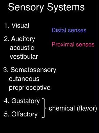
Sensory Systems. Picture: Rene Descartes (1596-1650). 1. Dr W Kolbinger, Sensory Systems (2009). Lecture Outline. Common Plan of Sensory Systems Four Sensory Receptor Classes Three Basic Processes in a Sensory Receptor Encoding of Four Stimulus Attributes

Dr W Kolbinger, Sensory Systems (2009)
E N D
Presentation Transcript
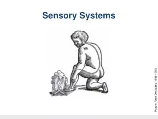
Sensory Systems Picture: Rene Descartes (1596-1650) 1 Dr W Kolbinger, Sensory Systems (2009)
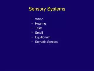
Lecture Outline • Common Plan of Sensory Systems • Four Sensory Receptor Classes • Three Basic Processes in a Sensory Receptor • Encoding of Four Stimulus Attributes • Convergence, Divergence and Lateral Inhibition 2
Common Plan of Sensory Systems Pathway Receptor Perception Behavior Stimulus Picture: Rene Descartes (1596–1650) 3
Fourth-order sensory afferent neurons Third-order sensory afferent neurons Second-order sensory afferent neurons First-order sensory afferent neurons Sensory receptors
Sensory Systems and their Receptors • Mechanoreceptors • Thermoreceptors • Chemoreceptors • Photoreceptors • Mechanoreceptors • Mechanoreceptors • Chemoreceptors • Chemoreceptors 5
Examples of Sensory Receptors • Sensory receptor neuron(somatosensory and olfactory systems • Sensory receptor cell(visual, taste, and auditory systems 6
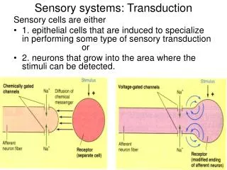
A B C D 1. Sensory receptors A: Free nerve endings (pain, temperature) B: Pacinian corpuscle (pressure) C: Meissner’s corpuscle (touch) D: Muscle spindle (stretch)
Three Basic Processes in Different Components of a Sensory Receptor • Cell body • transduction site • synaptic • Terminal • “graded potentials” 2) Action potentials 1) Receptor potential 3) Transmitter release 8
Encoding of Stimulus Intensity Sensory Transduction and Receptor Potentials 1. The environmental stimulus interacts with the sensory receptor and causes a change in its properties 2. receptor potential or generator potential. 3.receptor potentials are graded in amplitude
Encoding of Duration: Different Strategies • Slowly adapting receptors • remain active for the duration of a stimulus • Rapidly adapting receptors • are active only during times of changes (on/off) slowly adapting receptors are better in constantly monitoring levels of stimulation, whereas rapidly adapting receptors are most sensitive to changes, not to constant stimulation. 12
1 2 1 2 Encoding of Location:Receptive Field of a Sensory Receptor Stimulation Recordings A receptive field defines an area of the body that when stimulated results in a change in firing rate of a sensory neuron 13
Convergence and Divergence • Convergence • Divergence convergence answers the question “Where does the information come from?” divergence answers the question “Where does the information go to?” 14
lateral inhibition Receptive fields can be excitatory or inhibitory. The areas of inhibition contribute to a phenomenon called lateral inhibition, and aid in the precise localization of the stimulus by defining its boundaries and providing a contrasting border
Somatosensory System Lecture Outline • Five Modalities and their Receptors • Different Fibers for Different Receptors • Segmental Spinal Nerves and Dermatomes • Pathways for Different Modalities • Clinical Correlations 16
Modalities of the Somatosensory System • Touch (discriminative touch) • Vibration • Proprioception • Pain (nociception) • Temperature most of the somatic sensory modalities refer to sensations of the skin, proprioception refers to sensory receptors originating in the skeleto-muscular system. All receptors of the somatic sensory system are pseudo-unipolar neurons. Their sensory endings can be found in skin, or in (or close to) the muscle. Their fibers run in peripheral nerves and their cell bodies are located in ganglia (dorsal root ganglia in case the fibers run in spinal nerves or in cranial nerve ganglia). 17
Touch is transduced by Merkel’s disks (discriminative touch) and Ruffini’s endings (skin stretch). Vibration is transduced by Meissner’s corpuscles (for lower frequencies of about 50 Hz) and Pacinian corpuscles (for higher frequencies of about 300 Hz). Pain (pricking pain by rapidly adapting mechano-sensitive or thermo-sensitive receptors, burning pain by slowly adapting polymodal receptors) and Temperature (cold receptors and warm receptors) are transduced by free nerve endings.
Muscle spindles are embedded in extrafusal fibers of the working musculature of the muscle. The primary receptor of a muscle spindle is a rapidly adapting receptor with a Ia ("one a") afferent fiber. It carries sensory information of muscle stretch. This receptor forms the afferent limb of the myotatic reflex (deep tendon reflex), The secondary receptor of a muscle spindle is a slowly adapting receptor carrying sensory information of muscle length. It uses a class II ("two") afferent fiber. Golgi tendon organs are positioned close to the border between muscle and tendon. Their Ib afferent fibers form the afferent limb of the reverse myotatic reflex (inverse myotatic reflex).
Afferent Fiber Classification 12-20 μm 6-12 μm 1-6 μm 0.2-1μm A alpha A beta A delta C I II III IV 72-120 m/sec 36-72 m/sec 4-36 m/sec 0.4-2 m/sec 20
Afferent Fiber Classificationand Somatosensory Modalities 12-20 μm 6-12 μm 1-6 μm Touch, Vibration Proprioception Pain, Temperature 0.2-1μm • From skin: • From muscle: 21
Segmental Organization of Spinal Nerves and Dermatomes- segmental organization of the sensory innervation of the skin
Pathway for Touch, Vibration, Proprioception:Dorsal Column / Medial Lemniscus System Touch, vibration and proprioception are carried in a pathway called the dorsalcolumn / medial lemniscus system 23
Topographical Organization of the Spinal Cord • Dorsal column From arm From trunk From leg Midline 24
Spatial Orientation of Signals from Different Parts of the Body in Somatosensory Area • Somatosensory area has a high degree of localization • of the different parts of the body
Topographical Organization of the Spinal Cord • ALS From arm From trunk From leg Midline 28
Topographical Organization of the Spinal Cord Touch, vibration, proprioception from left side of body Pain, temperature from right side of body L T A Right Left L T A 29
Lissauer’s Tract and the Anterolateral System Right Left 30
Chief Complaint: Left Leg Paralysis, Right Leg Numbness History: A 75 year old retired pastry maker was in good health until about one year ago, when he started to develop gait difficulties and numbness of his right leg. Recently, he also experienced urinary urgency with occasional incontinence. Though he started taking laxatives, he experienced problems with bowel movements. His wife reported that he also had stiffness in the legs bilaterally. At times, the left leg has unexpectedly not been able to support his body weight for a brief period, causing him to stumble to maintain balance. He also reported that in addition to the numbness in the right leg, he also has a constant tingling feeling in the same limb, which he describes as “intolerable.” General Examination: Normal vital signs. Patient has no significant cardiovascular history. Abdomen was soft and non-tender. No palpable abdominal masses. Digital rectal examination showed significantly reduced muscle tone in the external sphincter and weakness of voluntary contraction. Prostate was felt to be enlarged with a highly nodular, irregular surface. Neurological examination: Patient was fully alert and oriented x 3. Cranial nerve exam was unremarkable. Upon motor examination, the upper extremities had normal strength, bulk, and tone, and the reflexes were 2+ throughout the C5 to C8 spinal level. In the lower extremities, however, the muscle tone was increased in left leg and the left iliopsoas muscle was weaker than the right (4/5). The muscle bulk in both legs was normal. Reflexes in the right leg were 2+, knee jerk on the left leg was 3+, and ankle jerk was 4+. Plantar response was extensor the left and flexor on the right. Finger to nose and heel to shin testing was normal. Pinprick testing and temperature sensation showed decreased sensitivity on the right side of the trunk below the umbilicus. Vibration and joint position sense was significantly reduced in the left leg and foot.
Brown Sequard Syndrome X X X X 32
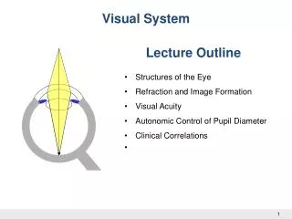
Visual System Lecture Outline • Structures of the Eye • Refraction and Image Formation • Visual Acuity • Autonomic Control of Pupil Diameter • Clinical Correlations 33 33 Dr W Kolbinger, Visual System (2009)
The Ocular Fundus Optic disc Fovea Macula The optic disc region itself only contains axons of retinal ganglion cells, the output elements of the retina, but it lacks photoreceptors. As a consequence, the optic disc is responsible for the blind spot, a region inside the boundaries of the visual field, where we don’t receive visual information. 35
Optics of the Eye Cornea refractive power: 42 D Flat lens refractive power: 13 D Rounded lens refractive power: 26 D Plasticity: 13 D 36
Accommodation Flat lens refractive power: 13 D • Far Vision Ciliary muscle relaxed Suspensory ligaments tightened Focus on the Retina Accommodation Adjusts the Refractive Power of the Eye 37
Accommodation Rounded lens refractive power: 26 D • Near Vision Ciliary muscle constricted Suspensory ligaments floppy Focus on the Retina 38
Presbyopia Flat lens • Near Vision The variability of the refractive power of the lens between far vision (13 D) and near vision (26 D) is called refractive plasticity. Unfortunately, the lens looses its elasticity during aging, thereby reducing the ability to focus on near objects, a condition called presbyopia. Blurred picture on the Retina 39