Download
1 / 71
750 likes | 1.1k Vues
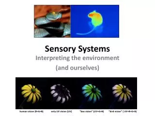
Sensory Systems. Environment-----> Sensing. Brain analysis. Action. organism. A view of animal behavior. What do sensory systems do?. Sensory receptors transduce the energy of stimuli and transmit signals to the central nervous systems.

E N D
Environment-----> Sensing Brain analysis Action organism A view of animal behavior
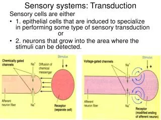
What do sensory systems do? Sensory receptors transduce the energy of stimuli and transmit signals to the central nervous systems. Stimulus---->Reception/Transduction---->Sensation/Perception Sensory Receptors CNS Sensations are action potentials that reach specific areas of the brain. Once the brain recognizes a sensation, it interprets it, giving the perception (color, taste, sound, taste) of a stimulus. Sensations and perceptions begin with sensory reception, which is the detection of a stimulus by sensory cells (or receptors)
Sensory receptors are specialized neurons. They can occur singly or in groups as part of sensory organs. Exteroreceptors are receptors that detect stimuli coming from outside of the body (heat, light, chemicals, pressure). Interoreceptors detect stimuli coming from within the body (blood pressure, plasma osmolality, blood pH).
-Sensory systems detect information from the environment (sensing), analyze it, and elicit actions. -Sensory receptors transduce the energy of stimuli and transmit signals to the central nervous systems. -Sensations are action potentials in response to specific stimuli that reach specific areas of the brain. Once the brain recognizes a sensation, it interprets it, giving the perception (color, taste, sound, taste) of a stimulus. -Exteroreceptors are receptors that detect stimuli coming from outside of the body (heat, light, chemicals, pressure). Interoreceptors detect stimuli coming from within the body (blood pressure, plasma osmolality, blood pH).
Receptors convert the energy of a stimulus into a change in their membrane potential. This change is graded (their magnitude depends on the magnitude of the stimulus) and is called a receptor potential. Receptor potentials result from the opening or closing of ion channels.
The receptor potential is transmitted either as a series of action potentials (their frequency depends on the magnitude of the receptor potential), or in the release of a neurotransmitter (the amount of neurotransmitter depends, again, on the magnitude of the receptor potential).
Sensory receptors have four functions: Sensory transduction (they transduce [transform/translate] the energy of a stimulus into a change in membrane potential). Amplification (they strengthen the energy of the stimulus. The action potential conducted from the eye to the brain contains 100,000 times more energy than the few photons of light that stimulated the receptor). Transmission (action potentials from receptors, or from neurons connected to receptors, reach the CNS). Integration (receptors contribute to the processing of a signal. For example many receptors show sensory adaptation. This term means that they respond less during continued stimulation. Of course some receptors “adapt” more quickly than others).
-Sensory receptors convert the energy of a stimulus into a graded change in their membrane potentialcalled a receptor potential. -Receptor potentials result from the opening or closing of ion channels. -A receptor potential is transmitted either as a series of action potentials or by the release of a neurotransmitter. -Sensory receptors have 4 functions: Sensory transduction (transform/translate the energy of a stimulus into a change in membrane potential), Amplification (they strengthen the energy of the stimulus). Transmission (action potentials from receptors, or from neurons connected to receptors, reach the CNS). Integration (receptors contribute to the processing of a signal).
Thermoreceptors (sense temperature) Pain receptors (nociceptors, from nocere (L) to hurt, usually have naked dendrites) Mechanoreceptors (sense physical deformation) There are many types of sensory receptors:
Chemoreceptors, include both receptors that transmit the total solute concentration of a solution (osmoreceptors), and specific receptors that respond to individual kinds of molecules. For example, many insects have gustatory hairs (sensilla) on their feet and mouthparts. Each sensillium contains 4 chemoreceptors, which respond differently to different chemical stimuli.
Animals also have Electromagnetic receptors which detect electromagnetic energy (including visible light, electricity, and magnetism)
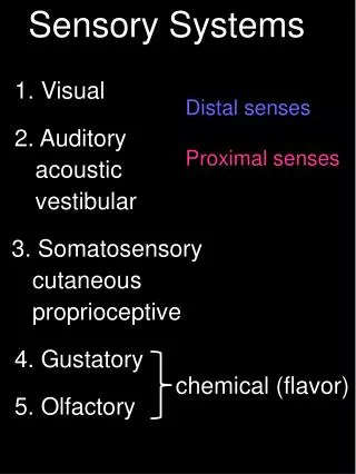
Types of Sensory Systems Hearing and Equilibrium (both rely on mechanoreceptors) Vision Taste and Smell (both rely on chemoreceptors) In many invertebrates, the position in which the statoliths (“position stones”) settle inside a statocyst (“position detector”) give the brain infoirmation about the orientation of the body. In humans, odorant molecules bind to specific receptors in the plasma membrane of osmoreceptors triggering action potentials.
Taste papillae 5 types of taste
-There are many types of receptors: thermoreceptors, nociceptors, mechanoreceptors, chemoreceptors (including osmoreceptors), electromagnetic receptors. -These receptors are found as elements of sensory systems including:hearing and equilibrium, taste and smell, and vision. -Both hearing and equilibrium depend on mechanoreceptors. Taste and smell depend on chemoreceptors. -Taste receptors can detect 5 types of “tastes”, salty, sweet, bitter, sour, and umami.
Vision Many types of “light detectors” have evolved among animals. These range from simple clusters of cells that detect the direction and intensity of light to complex organs that form images. In spite of great diversity, all photoreceptors contain similar pigment molecules that absorb light. Most of these pigments (opsins) are homologous (evolved from a common ancestral molecule). Planarians have two ocelli (light detecting organs, sometimes called eye spots or eye cups) that allow the animal to sense light (and often turn away from light). Orientation comes about by “comparing” light entering through each side. The cross-eyes in planaria serve a purpose!!
Two types of image-forming eyes have evolved among invertebrates. Compound eyes in arthropods (also some polychates) and single-lens eyes which have evolved in some polychaetes (annelids), in spiders, and in octopi and squid. Single-lens eye of squid Arthropod compound eye
Compound eyes are made of lots of ommatidia. The cornea and crystalline act as a lens that focuses light. Each ommatidium detects light on a narrow portion of the visual field and then the brain integrates this mosaic. Some insectscan detect color in the ultraviolet range. We cannot extrapolate our sensory world to other species. We Insect (maybe)
TO REMEMBER -Although animals use many types of light detectors (phtoreceptors), all of them use similar pigments (opsins). -Planaria have very simple ocelli (eye spots). -Invertebrates have ocelli, compound eyes (artropods, annelids), and single-lens eyes (octopi, squid). -Compound eyes are made of omatidia (cornea, crystalline, rhabdom, photoreceptor).. -Single-lens eyes of vertebrates have sclera, cornea, lens, iris, retina, optic nerve.
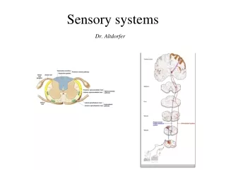
The retina contains photoreceptors (rods and cones), cells that integrate information across the retina (horizontal cells and amacrine cells), and cells that receive information from several rods and cones (bipolar cells) and relay it to ganglion cells which transmit action potentials to the brain by the optic nerve.
Each optic nerve has ≈ 106 axons that connects with interneurons in a structure called the geniculate nuclei. These interneurons relay sensations to the primary visual cortex, which is one of the brain centers responsible in constructing visual perceptions.
The photoreceptors in the retina of vertebrate eyes have two morphologies: they can be rod-like (“rods”) or cone-like (“cones”). Rods Cones
Rods are very good at detecting light at low intensities and at the low end of the electromagnetic spectrum, but are not good at distinguishing colors. Cones can detect variation in colors but are not as sensitive to light as rods. The reason for this difference is in the molecular structure of the pigments (opsins) contained in these two types of receptors. All vertebrate classes (fishes, amphibians, reptiles, and birds) have color vision. Most mammals are nocturnal and hence have mostly rods. Old World primates (including humans) are exceptional in that we have lots of cones (we will discuss the evolution of color vision a bit later). rod
In addition to the opsin found in rods (called rhodopsin), humans (and most Old World primates) have 3 different pigments which absorb (and hence are stimulated) at different peaks of the spectrum. For this reason, our visual system is called “trichromatic”
TO REMEMBER • The retina contains photoreceptors (rods and cones), cells that integrate information across the retina (horizontal cells and amacrine cells), and cells that receive information from several rods and cones (bipolar cells) and relay it to ganglion cells which transmit action potentials to the brain by the optic nerve. • -Each optic nerve has axons that connects with interneurons in a structure called the geniculate nuclei. These interneurons relay sensations to the primary visual cortex, which is one of the brain centers responsible in constructing visual perceptions. • -Rods are good at detecting light at low intensities but are not good at distinguishing colors. Cones can detect variation in colors but are not as sensitive to light as rods. • -Our visual system is called “trichromatic” because in addition to rhodopsin, we have 3 different pigments which absorb at different peaks of the spectrum.
Retinal exists in two forms. Light converts the cis form to a trans form and enzymes return it to its original form. Rods contain the visual pigment rhodopsin, which is embedded in a stack of membrane disks. Rhodopsin consists of the protein (pigment) opsin and the light absorbing molecule retinal (there are many opsins!).
The stimulus by which rods and cone stimulate or inhibit bipolar cells is the release of the neurotransmitter glutamate. + - To ganglion cells and then to brain by optic nerve.
Transducin activates PDE (phosphodiesterase) Light isomerizes retinal and activates rhodopsin The channels close and the membrane hyperpolarizes and stops releasing the neurotransmitter glutamate. Active rhodopsin Activates transducin Activated PDE detaches cGMP from Na+ channles in the plasma membrane.
TO REMEMBER • Rhodopsin, which is embedded in a stack of membrane disks. Rhodopsin consists of the protein (pigment) opsin and the light absorbing molecule retinal (there are many opsins). • Light starts a cascade in rhodopsin that leads to the closing of Na+ channels, the cell hyperpolarizes (its membrane potential becomes more negative) and stops releasing glutamate that stimulate a bipolar cell. The bipolar cell generates action potentials in the optic nerve. • The absence of light depolarizes the cell. The presence of light hyperpolarizes it.
Color vision, olfaction, fossil genes, and primate evolution Background: Olfactory receptor genes, which provide the basis for the sense of smell, represent the largest gene superfamily in mammalian genomes (> 1000 genes!!)
2) Trichromatic vision has evolved independently twice in primates. Both times as a result of opsin gene duplications, followed by mutations that modify the duplicated genes.
3) In the absence of natural selection, genes accumulate deleterious mutations that turn them into pseudogenes (“fossil genes”) that are not functional. Vision and smell seem to be alternative senses. In primates with trichromatic vision, a much higher % of olfactory receptor genes (30%) have become fossilized (pseudogenes) than in primates without trichromatic vision (≈ 15%).
Animal skeletons have three primary functions Support (most animals would sag against their own weight if they had no skeleton. We all fight gravity! Skeletons also preserve form) 2) Protection (a hard skeleton protects soft tissues. Think about skulls and ribs) 3) Movement (skeletons provide attachment sites for muscles, which are the engines for movement)
There are three main types of skeletons 1) Hydrostatic skeletons have fluid under pressure in a closed body compartment. Animals control movement by using muscles to change the shape of fluid-filled compartments. Hydrostatic skeletons are found in cnidarians, flatworms, and annelids. In earthworms, contraction of longitudinal muscles thickens and shortens segments of the worm; contraction of circular muscles constrict and elongate the segments. Bristles act as anchors.
Exoskeletons are deposited on the surface of the animal. Examples: The calcareous (made of calcite or calcium carbonate) shells of mollusks. 2) The jointed exoskeleton of arthropods and crustaceans are made of chitin and sometimes are reinforced by calcite and other materials.
The exoskeleton of arthropods is secreted by the epidermis (a layer of living cells) and it is made of chitin and protein. In insects it is covered by a layer of impermeable wax, which reduces evaporation.
Endoskeletons consist of hard supporting elements (such as bones), buried within the animal’s soft tissues. Chordates have skeletons made of cartilage (mostly collagen) and bone. Echinoderms have endoskeletons under made of hard plates called ossicles under their skin. The ossicles are made of magnesium and calcium carbonate bound by protein fibers.
The skeleton of mammals has over 200 bones (some of you will have to learn all their names one day!), some fused, some connected at joins by ligaments. Anatomists divide the vertebrate skeleton into two main parts: the axial skeleton (skull, vertebral column, and rib cage), and the appendicular skeleton (limbs, and pectoral and pelvic girdles).
Three types of joints Enable us to rotate our arms and legs (where the femur contacts the pelvic girdle) and move them in several planes. Restrict movements to a single plane Allow rotation of our forearm at the elbow (and our head from side to side).
Animals can move as a result of the action of muscles Muscles act by contracting. The ability to move parts of the body in opposite direction requires that different muscles are attached to the skeleton in “antagonic pairs” that work against each other.
The structure of vertebrate skeletal (striated) muscle Vertebrate skeletal muscle attaches to bones and is responsible for their movement. It is characterized by a hierarchy of smaller and smaller units. 1) Each muscle is a bundle of muscle fibers. 2) Each muscle fiber is a single multinucleated cell and is a bundle of many myofibrils. 3) The myofibrils, in turn, are made of two kinds of myofilaments. 4) Thin filaments made of actin and a regulatory protein, and 5) Thick filaments , which are arrays of the protein myosin. The regular arrays of filaments give skeletal muscle as striated appearance.
I bands and H zone are relatively wide The sliding-filament model of muscle contraction is our best hypothesis to explain how muscle contracts. Note that the length of the thick and thin filaments remains the same as the muscle contracts. However, the length of the sarcomer is shorter. Thick and thin filaments slide past each other. The width of the I bands and and H zone is reduced