Download
1 / 51
510 likes | 1.74k Vues
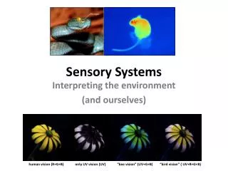
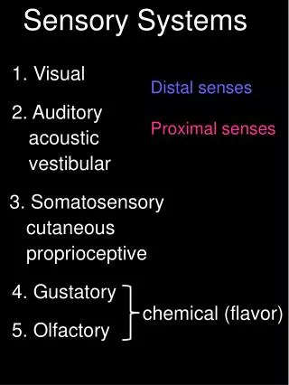
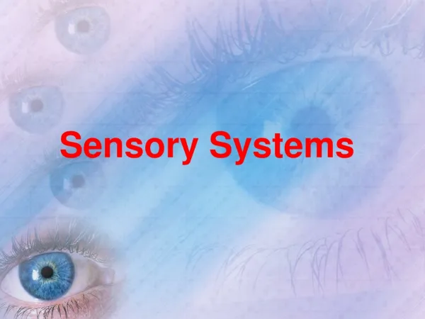
Sensory Systems. Tongue taste. Ear Hearing. Eye sight. Skin touch. Sensory Receptors. Sensory receptors are cells that capture all information about the environment that is processed by the brain . Receptor cells are an integral parts of sensory organs which are:. Nose

E N D
Tongue taste Ear Hearing Eye sight Skin touch Sensory Receptors • Sensory receptors are cells that capture all information about the environment that is processed by the brain. • Receptor cells are an integral parts of sensory organs which are: Nose smell
Balance!!! • Some receptor cells are also in muscles to be aware of the stretching, as well as in the inner ear for balance. • Combined with sense of sight, receptors in muscles and the inner ear allow the body to hold a position or move, this also involves a sense of balance and orientation.
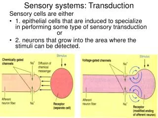
Sensory Messages • Each of the sensory organs sends messages through the nervous system. • These messages are interpreted by the brain. • A sensory message always sends a signal along the same path. 1. Sensory receptor captures information (stimulus) 2. A “transformer “ changes info into a nerve impulse 3. Sensory neurons send info to the brain to be analyzed.
When our brain does not understand the signals it is sent, it tries to make sense of them • Ex: Optical illusions
The Eye (sight) The eye is a complex, sensory organ that is sensitive to light. Sphere-shaped, 2.5cm in diameter The eyelids, eyelashes and eyebrows protect the eyes.
The Anatomy of the Eye • The eye is made up of the: • Cornea • Sclera • Iris • Lens • Vitreous Gel • Retina • Macula • Optic nerve Sclera
Parts of the eye • The eye has three opaque membranes (light cannot pass): • Thesclera • The choroid • The retina
The transparent parts of the eye allow light to pass through the eye and converge on the retina. • These are in order: • The cornea • The aqueous humor • The lens • The vitreous humor
a lens Vitreous humor cornea Aqueous humor
Sclera • Rigid, thick, opaque membrane, 1 mm thick, “white of the eye”. • Outside layer which protects the eye from shock and gives it its distinctive shape; • The thick and opaque layer of conjunctive tissue becomes thinner and transparent at the front of the eye, where it bulges slightly, to form the cornea
Choroid • Middle layer of the eye. • It is dark brown and has many blood vessels which supplies nutrients and oxygen to the different layers of the eye • In the front of the eye, the choroid becomes the iris.
Iris Pupil: the centre of the iris where light enters • The iris is the coloured part of the eye. • Composed of two groups of muscles, in the centre, which controls the amount of light that can enter the eye and reach the retina.
The pupil is the opening formed by the iris • The iris dilates(widens) and the pupil contracts when in bright light, allowing less light to enter the eye. • The iris contracts and becomes narrower and the pupil widens when it is dark, allowing more light to pass through.
Retina • Pinkish-beigecolored membrane. • This is the lining at the back of the eye composed of specialized nerve cells . • The Retina covers 2/3 of the inside of the eye and ends in a jagged edge. • This makes it susceptible to detaching
The cells in the retina are sensory receptors and are photosensitive (they react to light). • They are called photoreceptors and change light into electrical impulses. • They are stimulated when light rays enter your eyes after bouncing off objects in surroundings.
There are two types of photoreceptor cells in the retina: *Rods *ConesRetina can turn light into nerve impulse using the cones and rods that are connected to sensory neurons
Cones: • There are about 20x less cones than rods • They are concentrated in the center of the retina in a spot (a cone filled pit) approx2mm in diameter called the macula. MACULA • This is at the centre of the retina. • It is where light is focused for central vision. • Most cone cells are found at the centre of the macula
The retina The macula
Cones are responsible for color and very detailed vision but need lots of bright light for this. (you see less color in detail when it gets dark) • There are 3 types of cones: • Red – detect red • Green – detect green • Blue – detect blue • Red and some green = orange • All three stimulated equally results in white color
Rods • These cells are responsible for peripheral vision (what we see to the sides, above and below what we are looking at). • Rods detect contrast, form and shape of objects • Rods are important for night vision-dim light(seeing in the dark) • No rods in macula, as you get further from macula, there are more rods. • Rods cover the retina except in macula and the “ blind spot”
Optic Nerve • The optic nerve is the bundle of nerve cells that passes the information from the retina to the brain. • There are no photoreceptors here so we get a “blind spot”
The junction point between the optic nerve and retina, the BLIND SPOT, has NO sensitivity to light
The optic disc is the part where there are no cones or rods and it is where the optic nerve begins.
Cornea • Clear and rigid membrane that is an extension of the sclera and covers the front of the eyeball where light passes through • The muscles around the eye help the cornea to change shape and helps us focus. The cornea does about 2:3 of the eyes focusing. • It is slightly domed shaped which allows it to converge light rays; • If the cornea is irregular in shape or has malformations, it can lead to vision problems. (myopia and astigmatism)
Aqueous humour • Transparent liquid that fills the space between the iris and the cornea; • Consists of water (which is always replenished) and a little glucose; • It supplies glucose and oxygen to the lens, cornea and cells around the retina and it also eliminates waste • Allows light rays to travel through it
Lens • It is behind the iris; • It is flexible; • It has a biconvex shape (convex on both sides) • Approx 9mm dia. and changes its shape from 4-5mm thick depending on whether you are looking at a far object or a close one.
Lens Continued….. • A healthy lens is completely transparent; • The Lens brings the image that enters the eye and projects onto the retinainto clear focus • It gets thicker (due to the action of ciliary muscles and zonular ligaments) to see nearby objects. It relaxes and returns to its less curved form to see distant objects. This is called accommodation. • Thus the lens can move inside the eye (stretching and contracting) to let us focus on near and far things.
Cataracts happen when the lens becomes opaque (turns cloudy) • With time the lens becomes less flexible which leads to presbyopia and requires corrective lenses/glasses.
Vitreous Humour • This is a thick, transparent, jelly-like substance mainly composed of water, fills the eye between the lens and the retina. Lets light rays pass through to the retina. • It occupies the largest part of the eye • Main function: Helps the eye keep it’s shape because: • it exerts pressure on the membranes to keep the shape of the eye and keep the lens and retina in place; • It eliminates waste from the lens and retina and supplies them with glucose and oxygen. It is constantly renewed.
iris sclera choroid retina Optic nerve Vitreous humor lens Aqueous humor cornea
Auxiliary structures • The eye is surrounded by various structures that aid in its protection • The eyebrow • Keeps sweat from going into the eyes • The eyelashes • Keep dust out
The eyelids • wipe the eye and spread the tears over the eye • The tear(lacrimal) glands • produce tears that clean and lubricate the eye • Tears are antibacterial • Tears contain water and mineral salts • Tears wash across the eye and excess liquid drains into the lachrymal duct (tear duct) leading to the nose (this is why your nose runs when you cry)
Physiology of the Eye • Transparent environments in the eye transmit light rays to the retina, where the nerve cells turn the light rays into nerve impulses. • The impulses travel through the optic nerve to the visual area of the brain, which analyzes and interprets them. • The brain superimposes (copies) the image it received from each eye • This is how we are able to estimate distances and objects contours (shapes)
How the Eye Sees…when light travels through a dense transparent substance with different curvature, it is deviated. 1. Converging lenses: cause light rays to meet, the light rays come together 2. Diverging lenses: cause light rays to spread out, The concave lens makes light deviate/diverge Focal point Focal point
Lens accommodation • When looking at a distant object the incoming light is parallel • so the lens does not have to change its shape. Rays converge and focus image on the retina *
When looking at close objects the light rays from the object are diverging (move away from one another) • so the lens must become “fatter” in order to make them converge and focus the light on the retina
Lens accommodation • The accommodations are possible because of the contractions of small ciliary muscles that pull on the zonular ligaments attached to the lens; • An image is projected on the retina each time light passes through the transparent layers of the eye but it is inverted by the lens(as it would be with any biconvex lens) and will be interpreted normally by the brain.
Nerve impulse pathways • Light penetrates the eye, strikes the cones and rods (processors) of the retina and they become stimulated and then turn the light rays into a signal that produces nerve impulses; • The impulses are transmitted by the sensory neuron and travel along the axon in the optic nerve (conductor); • The nerve extends to the visual area of the brain (analyzer) at the back of the head. The sensation of seeing does not occur until the brain has analyzed the info
Correcting eye problems with lenses • Emmetropia: normal state of the eye when it is able to focus light properly on the retina producing a clear image, if the image is blurred, there is an eye problem such as the following: • Presbyopia:. makes nearby objects difficult to see. Is caused by a loss of lens flexibility associated with aging. The problem is cured with a biconvex lens (bi-focals) • Astigmatism: makes it hard to see both distant and nearby objects. Caused by an irregularity in the curvature of the cornea or lens (shaped like a football). The light rays passing through these transparent environment deviate in different directions and project a blurred image onto the retina. It can be cured with cylindrical lenses that concentrate light rays and limit deviation.
Myopia: near-sightedness • Can see close but not far away • Occurs when the cornea is too curved or the eye is too long. • The light rays converge before reaching the retina and the clear image ends up in front of the retina • The one reaching the retina is blurred and that is the one that is analyzed by the brain. • Fix it with a concave(diverging) lens (make the light rays diverge until they reach the lens, then converge on the retina)
Hyperopia: far-sightedness • See distant objects well, but close up objects blurry. • It is caused by a slightly shortened eyeball or an cornea that is too flat. • The image is focused past the retina and appears blurred • The problem can be corrected with a biconvex lens, which will make the light rays converge on the retina.
Hyperopia Convex lens for correction
Cataracts: Caused by a lens that gradually becomes opaque, prevents light from reaching retina. Occur at about 65 yrs old causes include diabetes or other metabolic diseases, smoking, uv rays, or trauma.