Sensory Systems
Sensory Systems. Christopher Skinner MD FRCPC Ottawa Hospital cskinner@toh.on.ca. Sensory Systems. Objectives. 4799 Describe the anatomical features of the somatosensory pathways. 4800 Explain the effects of damage to these pathways at different locations .

Sensory Systems
E N D
Presentation Transcript
Sensory Systems Christopher Skinner MD FRCPC Ottawa Hospital cskinner@toh.on.ca
Sensory Systems Objectives • 4799 Describe the anatomical features of the somatosensory pathways. • 4800 Explain the effects of damage to these pathways at different locations. • 4801 Describe in general terms the mechanisms for generation of electrical signals by sensory stimuli (sensory transduction). • 4802 Discuss the means by which the characteristics of sensory stimuli are encoded by signals to the cerebral cortex. • 4803 Define receptive fields of neurons. • 4804 Briefly explain the effects of lesions at peripheral vs. central levels of the auditory pathway. • 4999 Compare visual pathway anatomy to other sensory pathways. • 5000 Describe the anatomical basis of hemineglect. • 5470 Describe the basic characteristics of hemineglect.
Sensory Systems • Lecture Outline • Sensory receptors and transduction • mechanoreceptors • G-protein-coupled receptors • Sensory coding • Receptive fields • lateral inhibition and contrast enhancement • Somatosensory pathways • Effects of lesions • Visual pathways • Effects of lesions • The pupillary light reflex • Sensory association areas of the cortex • Hemineglect • Averaged evoked potentials
Somatosensory Receptors Touch Receptors Primary Afferent fibre Meissner’s Merkel Ruffini Pacinian Receptive fields Action potentials Phasic/tonic Stimulus
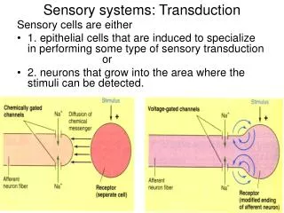
Sodium influx Potassium efflux Local Anesthetic SensoryTransduction 1 msec Local anesthetics block voltage-sensitive sodium channels, and prevent transmission of the action potentials along the nerve.
Saltatory Conduction • Depolarization at one node of Ranvier is sufficient to elevate the voltage at a neighboring node. • The voltage at the first node of Ranvier extends spatially to the next node of Ranvier. • At each successive node, the membrane potential of the axon is thereby brought to the threshold potential to initiate an action potential. • Ions need only to cross the axon membrane to propagate the action potential at the nodes, but not anywhere under the myelin along the axon. • In myelinated axons, action potentials do not propagate continuously as waves, but instead recur at successive nodes, and in effect "hop" along the axon, by which process they travel faster than they would otherwise. • In summary, the charge will passively depolarize the adjacent node of Ranvier to threshold, triggering an action potential in this region and subsequently depolarizing the next node, and so on. Wikipedia
Stretch-sensitive Channels in Mechanoreceptors Deformation of the sensory nerve terminal membrane changes the ion permeability of the stretch-sensitive channels. For the somatosensory system, this always leads to depolarization of the membrane.
A Puzzle! • Recall that action potentials are all-or-none electrical signals that are very similar i.e. binary events. • How, then, does the CNS know the nature and strength of the stimulus that gave rise to these signals?
Coding of Sensory Signals Slowly Adapting Receptor Rapidly Adapting Receptor Action Potentials in Sensory Nerve (appear as lines because of slow time base) Threshold for AP Receptor Potential Stimulus (1 second) The duration and strength of a stimulus can be coded by the duration and frequency of the resulting train of action potentials, respectively.
Vestibular Hair Cell In auditory and vestibular hair cells, movement of the hairs (by vibration, or motion of the surrounding fluid) opens ion channels. The resulting electrical changes can be transmitted chemically to the associated nerve terminals and converted to action potentials that send the sensory signal to the CNS.
Luminance (logarithmic scale) -6 -4 -2 0 2 4 6 8 Indoor lighting sunlight starlight moonlight Cone threshold Absolute threshold Rod saturation Possible damage Broad-range Sensitivity to Stimulus Intensity (No colour vision)
Receptive Fields • Receptive fields of single nerve cells can be described by the region that, when stimulated, will produce an electrical response in that cell.
Receptive Fields for Retinal Ganglion Cells On-centre Cell Off-centre Cell Inhibitory centre Excitatory centre Excitatory surround Inhibitory surround Light On Light On Centre Illuminated Surround Illuminated Diffuse Illumination
Neuronal Circuitry in the Retina • Lateral inhibition! • A mechanism to enhance • contrast between signals that • are spatially close. • This permits better discrimination • of the borders of regions of stimulation. Recording electrode + - + Note that only the ganglion cell on the left is being recorded, so the effect of light stimulation on the right is inhibitory (i.e. in the “surround” Part of the receptive field) - + +
Lateral Inhibition in the Somatosensory System Improved discrimination
Somatosensory Systems Touch receptors Posterior Column System Anterolateral System
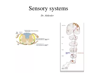
Schema of Somatosensory Pathways Cortex Thalamus Brain Stem (medulla) Spinal Cord Trigeminal Systems Posterior column pathway Spinothalamic pathway (anterolateral) Somatosensory Pathways
Why do the amounts of white matter and grey matter vary at different levels of the spinal cord?
Topographic Arrangement of Somatosensory Pathways Posterior columns (disc. touch, vib’n, proprioception) S L T Cortico-spinal tract (motor) C sensory S L T Spinal grey C C T L S motor Spino-thalamic tract (pain, temp., touch)
Topographic Arrangement of Somatosensory Pathways Posterior columns (disc. touch, vib’n, proprioception) S L T Cortico-spinal tract (motor) C sensory S L T Sacral Sparing Spinal grey C C T L S motor Spino-thalamic tract (pain, temp., touch)
The area of somatosensory cortex representing a given part of the body is related to the number of sensory receptors innervating that area. • Activation of cortical neurons in a primary sensory area initiates the perception process. • Normally, the activation is through the sensory path, and the modality and location of the stimulus are indicated by the cortical region, and location within that region, of the activated neurons.
Lesionsof Somatosensory Pathways Cortex Thalamus Brain Stem (medulla) Spinal Cord Trigeminal Systems Posterior column pathway Spinothalamic pathway (anterolateral) Somatosensory Pathways
Cerebral Arterial Supply Because of functional localization within the cortical areas, localized vascular occlusions can give different clinical signs.
Brainstem Vascular Lesions Regional ischemia due to vascular lesions in other areas may also give differential clinical signs.
Spino-cerebellar Pathways Friedreich’s Ataxia, an autosomal recessive disorder, is associated with fewer large sensory afferents and reduced spino-cerebellar and dorsal column pathways.