Sensory Systems
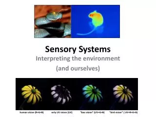
Sensory Systems. Vision Hearing Taste Smell Equilibrium Somatic Senses. Sensory Systems. Somatic sensory General – transmit impulses from skin, skeletal muscles, and joints Special s enses - hearing, balance, vision Visceral sensory Transmit impulses from visceral organs

Sensory Systems
E N D
Presentation Transcript
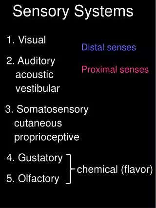
Sensory Systems • Vision • Hearing • Taste • Smell • Equilibrium • Somatic Senses
Sensory Systems • Somatic sensory • General – transmit impulses from skin, skeletal muscles, and joints • Special senses - hearing, balance, vision • Visceral sensory • Transmit impulses from visceral organs • Special senses - olfaction (smell), gustation (taste)
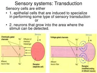
Properties of Sensory Systems • Stimulus - energy source • Internal • External • Receptors • Sense organs - structures specialized to respond to stimuli • Transducers - stimulus energy converted into action potentials • Conduction • Afferent pathway • Nerve impulses to the CNS • Translation • CNS integration and information processing • Sensation and perception – your reality
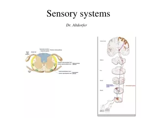
Sensory Pathways • Stimulus as physical energy sensory receptor acts as a transducer • Stimulus > threshold action potential to CNS • Integration in CNS cerebral cortex or acted on subconsciously
Classification by Function (Stimuli) • Mechanoreceptors – respond to touch, pressure, vibration, stretch, and itch • Thermoreceptors – sensitive to changes in temperature • Photoreceptors – respond to light energy (e.g., retina) • Chemoreceptors – respond to chemicals (e.g., smell, taste, changes in blood chemistry) • Nociceptors – sensitive to pain-causing stimuli • Osmoreceptors – detect changes in concentration of solutes, osmotic activity • Baroreceptors – detect changes in fluid pressure
Classification by Location • Exteroceptors – sensitive to stimuli arising from outside the body • Located at or near body surfaces • Include receptors for touch, pressure, pain, and temperature • Interoceptors – (visceroceptors) receive stimuli from internal viscera • Monitor a variety of stimuli • Proprioceptors – monitor degree of stretch • Located in musculoskeletal organs
Somatic Senses • General somatic – include touch, pain, vibration, pressure, temperature • Proprioceptive – detect stretch in tendons and muscle provide information on body position, orientation and movement of body in space
Somatic Receptors • Divided into two groups • Free or Unencapsulated nerve endings • Encapsulated nerve endings - consist of one or more neural end fibers enclosed in connective tissue
Free Nerve Endings • Abundant in epithelia and underlying connective tissue • Nociceptors - respond to pain • Thermoreceptors - respond to temperature • Two specialized types of free nerve endings • Merkel discs – lie in the epidermis, slowly adapting receptors for light touch • Hair follicle receptors – Rapidly adapting receptors that wrap around hair follicles
Encapsulated Nerve Endings • Meissner’s corpuscles • Spiraling nerve ending surrounded by Schwann cells • Occur in the dermal papillae of hairless areas of the skin • Rapidly adapting receptors for discriminative touch • Pacinian corpuscles • Single nerve ending surrounded by layers of flattened Schwann cells • Occur in the hypodermis • Sensitive to deep pressure – rapidly adapting receptors • Ruffini’s corpuscles • Located in the dermis and respond to pressure • Monitor continuous pressure on the skin – adapt slowly
Encapsulated Nerve Endings - Proprioceptors • Monitor stretch in locomotory organs • Three types of proprioceptors • Muscle spindles – monitors the changing length of a muscle, imbedded in the perimysium between muscle fascicles • Golgi tendon organs – located near the muscle-tendon junction, monitor tension within tendons • Joint kinesthetic receptors - sensory nerve endings within the joint capsules, sense pressure and position
Special Senses • Smell • Taste • Vision • Hearing • Equilibrium Figure 10-4: Sensory pathways
External Structures of the Eye Figure 17.3a, b
Eye anatomy • Ciliary body and lens divide the eye into posterior (vitreous) cavity and anterior cavity • Anterior cavity further divided into anterior and posterior chambers • Aqueous humor circulates within the eye • diffuses through the walls of anterior chamber • re-enters circulation • Vitreous humor fills the posterior cavity. • Not recycled – permanent fluid
The Pupillary Muscles Figure 17.5
Sectional Anatomy of the Eye • Outer fibrous tunic -sclera, cornea, • Vascular tunic - iris, ciliary body, choroid • Nervous tunic - retina Figure 17.4a, b
Organization of the Retina Figure 17.6b, c
Organization of the Retina Figure 17.6a
Retina Figure 10-38: Photoreceptors: rods and cones
Retina • Rod cells • Monochromatic • Night vision • Cone cells: • Red, green, & blue • Color & details • Pigmented epithelium • Melanin granules • Prevents reflection • Bipolar & ganglion cells converge, integrate APs
Vision: Photoreceptors • Reflected light translated into mental image • Pupil limits light, lens focuses light • Retinal rods and cones are photoreceptors Figure 10-36: Photoreceptors in the fovea
Lens – Image Formation • Lens helps focus • Light is refracted as it passes through lens • Accommodation is the process by which the lens adjusts to focus images • Normal visual acuity is 20/20
Accommodation Figure 17.10
Visual Abnormalities Figure 17.11
Photoreception and Local Integration Figure 10-35: ANATOMY SUMMARY: The Retina
Photoreception Retinal Changes Shape Retinal restored Opsin inactivated Figure 17.15
Convergence and Ganglion Cell Function Figure 17.18
Visual Pathways Figure 17.19
Equilibrium and Hearing Both Equilibrium And Hearing Are Provided By Receptors Of The Inner Ear
Middle Ear Figure 17.21
Inner ear • Membranous labyrinth contains endolymph • Bony labyrinth surrounds and protects membranous labyrinth • Vestibule • Semicircular canals • Cochlea
Cochlea Figure 17.25a, b
Sound and Hearing • Sound waves travel toward tympanic membrane, which vibrates • Auditory ossicles conduct the vibration into the inner ear • Movement at the oval window applies pressure to the perilymph of the cochlear duct • Pressure waves distort basilar membrane • Hair cells of the Organ of Corti are pushed against the tectoral membrane Figure 17.28a
The Organ Of Corti Figure 17.26a, b
Semicircular Canals • Provide information about rotational acceleration. • Project in 3 different planes. • Each canal contains a semicircular duct. • At the base is the crista ampullaris, where sensory hair cells are located. • Hair cell processes are embedded in the cupula. • Endolymph provides inertia so that the sensory processes will bend in direction opposite to the angular acceleration.
Utricle and Saccule • Utricle: • More sensitive to horizontal acceleration. • During forward acceleration, otolithic membrane lags behind hair cells, so hairs pushed backward. • Saccule: • More sensitive to vertical acceleration. • Hairs pushed upward when person descends.
Olfactory organs • Contain olfactory epithelium with olfactory receptors, supporting cells, basal cells • Olfactory receptors are modified neurons • Surfaces are coated with secretions from olfactory glands • Olfactory reception involved detecting dissolved chemicals as they interact with odorant binding proteins
Olfaction • Olfactory pathways • No synapse in the thalamus for arriving information • Olfactory discrimination • Can distinguish thousands of chemical stimuli • CNS interprets smells by pattern of receptor activity • Olfactory receptor population shows considerable turnover • Number of receptors declines with age
Taste Receptors • Clustered in taste buds • Associated with lingual papillae • Taste buds • Contain basal cells which appear to be stem cells • Gustatory cells extend taste hairs through a narrow taste pore
Gustatory pathways • Taste buds are monitored by cranial nerves • Synapse within the solitary nucleus of the medulla oblongata • Then on to the thalamus and the primary sensory cortex • Primary taste sensations • Sweet, sour, salty, bitter • Receptors also exist for umami and water • Taste sensitivity shows significant individual differences, some of which are inherited • The number of taste buds declines with age