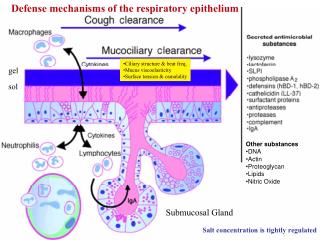
Submucosal Gland
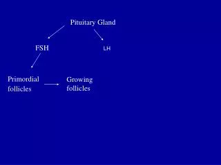
Defense mechanisms of the respiratory epithelium. Ciliary structure & beat freq. Mucus viscoelasticity Surface tension & osmalality. gel. sol. Other substances DNA Actin Proteoglycan Lipids Nitric Oxide. Submucosal Gland. Salt concentration is tightly regulated.

Submucosal Gland
E N D
Presentation Transcript
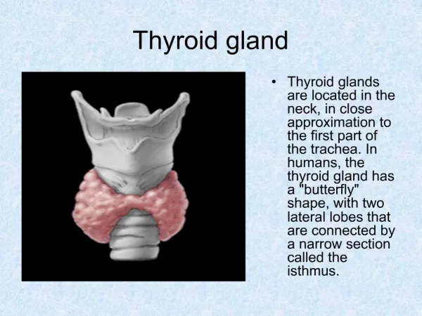
Defense mechanisms of the respiratory epithelium • Ciliary structure & beat freq. • Mucus viscoelasticity • Surface tension & osmalality gel sol • Other substances • DNA • Actin • Proteoglycan • Lipids • Nitric Oxide Submucosal Gland Salt concentration is tightly regulated
Apical Surface of Normal Airway Epithelium From Cell, 85(1996), 229
NK Cells Neutrophils (PMN) Macrophages Dendritic Cells Mast Cells Basophils Eosinophils Antimicrobial substances Inorganic disinfectants Small AMP Large AMP Complement Cytokines Innate Immunity is Comprised of Cellular and Humoral Factors
PHAGOCYTES MONOCYTE NEUTROPHILS Can discriminate between "foreign" and "self" molecules because they possess receptors for carbohydrates that are not normally exposed on the cells of vertebrates, such as mannose. In addition, both macrophages and neutrophils have receptors for antibodies and complement, so that the coating of microorganisms with antibodies, complement, or both enhances phagocytosis.
PhagocytosisBound pathogens are internalized in phagosome, which become acidified (pH 3.5 - 4.0).Lysosomes contain enzymes, proteins, and peptides that can mediate intracellular antimicrobial response.Phagosome fuses with one or more lysosomes to form a phagolysosome, in which lysosomal contents are released to destroy the pathogen.Macrophages that have bound and ingested microorganisms also contribute to the adaptive immune response by acting as antigen-presenting cells.
Neutrophil degranulation of antimicrobial proteins and peptides Specific (secondary) granules are more prone to degranulate their contents (including lactoferrin and cathelicidins) into the extracellular space. In contrast, azurophil (primary) granules, containing BPI and defensins, are predominantly degranulated into the phagolysosome. To a lesser extent, specific granules also degranulate into the phagolysosome and primary granules to the extracellular space.
Respiratory Burst Generated by Lysosomal NADPH oxidases Myeloperoxidase- independent Myeloperoxidase- dependent
Inflammatory Responses • Activated macrophages also produce cytokines and other mediators to trigger inflammatory responses. • Inflammation can also be triggered by the Complement Cascade. • Inflammation is traditionally defined by 4 Latin words, rubor, calor, tumor and dolor, meaning redness, heat, swelling and pain. • Inflammation plays three essential roles in combating infection: • deliver additional effector molecules and cells to sites of infection to augment the killing of invading microorganisms by the front-line macrophages, • provide a physical barrier preventing the spread of infection, and • promote the repair of injured tissue. • Reflects 3 types of changes in the local blood vessel: • an increase in vascular diameter, leading to increased local blood flow and a reduction in the velocity of blood flow, • endothelial cells lining the blood vessel are activated to express adhesion molecules that promote the binding of circulating leukocytes, and • increase in vascular permeability of local blood vessels, leading to exit of fluid and protein from the blood and their accumulation in the tissues.
Neutrophil Recruitment: the Multistep Model ICAM-1 GlyCAM-1/CD34/ MadCAM-1
ICAM-1 P-Selectin E-selectin Neutrophil Recruitment: the Multistep Model Sialyl-Lewis X PSGL-1 Sialyl-Lewis X
Dendritic cells that recognize pathogens, migrate out of infected areas and into nearby lymph nodes where fragments of pathogen are displayed to T cells. NEJM 343, 40(00)
The Complement System • The complement system is a set of plasma proteins that act together to attack extracellular forms of pathogens. • It was first discovered as an effector arm of the antibody response, but complement can also be activated early in infection in the absence of antibodies; complement first evolved as part of the innate immune system. • Activation of complement involves the sequential proteolysis of proteins to generate enzymes with proteolytic activity. • The products of complement activation become covalently attached to microbial cell surfaces or to Ab bound to microbes and to other antigens. • Complement activation is inhibited by regulatory proteins that are present on normal host cells and absent from microbes. • There are three pathways of complement activation: • the classical pathway, which is triggered by antibody or by direct binding of complement component C1q to the pathogen surface; • the MB-lectin pathway, which is triggered by mannan-binding lectin, a normal serum constituent that binds some encapsulated bacteria; and • the alternative pathway, which is triggered directly on pathogen surfaces. • The 3 pathways converge at the point of cleavage of C3.
The Three Activation Pathways of Complement: the Classical, Mannose-Binding Lectin, and Alternative Pathways. C9
The Alternative Pathway: Regulation of the Cleavage of C3. In fluid phase, C3b is inactivated by hydrolysis.