Ch 19
Blood Vessels. Ch 19. Blood Vessels. Blood is carried in a closed system of vessels that begins and ends at the heart The three major types of vessels are arteries, capillaries, and veins Arteries carry blood away from the heart, veins carry blood toward the heart

Ch 19
E N D
Presentation Transcript
Blood Vessels Ch 19
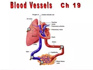
Blood Vessels • Blood is carried in a closed system of vessels that begins and ends at the heart • The three major types of vessels are arteries, capillaries, and veins • Arteries carry blood away from the heart, veins carry blood toward the heart • Capillaries contact tissue cells and directly serve cellular needs
Tunics • Tunica interna (tunica intima) • Endothelial layer that lines the lumen of all vessels • In vessels larger than 1 mm, a subendothelial connective tissue basement membrane is present • Tunica media • Smooth muscle and elastic fiber layer, regulated by sympathetic nervous system • Controls vasoconstriction/vasodilation of vessels
Tunics • Tunica externa (tunica adventitia) • Collagen fibers that protect and reinforce vessels • Larger vessels contain vasa vasorum
Blood Vessel Structure simple squamous epithelium smooth muscle tissue connective tissue
Tunica intima Valve • Endothelium • Subendothelial layer Internal elastic lamina Tunica media (smooth muscle and elastic fibers) External elastic lamina Tunica externa (collagen fibers) Lumen Vein Lumen Artery Capillary network Basement membrane Endothelial cells Capillary (b) Figure 19.1b
Artery vein
Arteries 1. Carry blood away from the heart. 2. Thick-walled to withstand hydrostatic pressure of the blood during ventricular systole. 3. Blood pressure pushes blood through the vessel
Elastic (Conducting) Arteries • Thick-walled arteries near the heart; the aorta and its major branches • Large lumen allow low-resistance conduction of blood • Contain elastin in all three tunics • Withstand and smooth out large blood pressure fluctuations • Allow blood to flow fairly continuously through the body
Muscular (Distributing) Arteries and Arterioles • Muscular arteries – distal to elastic arteries; deliver blood to body organs • Have thick tunica media with more smooth muscle and less elastic tissue • Active in vasoconstriction • Arterioles – smallest arteries; lead to capillary beds • Control flow into capillary beds via vasodilation and constriction
Fig.21.02 Pressure reservoir function of elastic arteries
Veins • Carry blood to the heart. • Thinner-walled than arteries. • Possess one-way valves that prevent backwards flow of blood. • Blood flow due to body movements, not from blood pressure.
One-Way Valves in Veins open valve closed valve
Capillaries • One cell thick, endothilium • Function: Diffusion Filtration Transcytosis
Diffusion O2 CO2
Transcytosis Lumen Caveolae Pinocytotic vesicles Endothelial fenestration (pore) Intercellular cleft 4 Transport via vesicles or caveolae (large substances) 3 Movement through fenestrations (water-soluble substances) Basement membrane 2 Movement through intercellular clefts (water-soluble substances) 1 Diffusion through membrane (lipid-soluble substances) Figure 19.16 (2 of 2)
Capillaries • Capillaries are the smallest blood vessels • Walls consisting of a thin tunica interna, one cell thick • Allow only a single RBC to pass at a time • Pericytes on the outer surface stabilize their walls • There are three structural types of capillaries: continuous, fenestrated, and sinusoids
Continuous Capillaries • Continuous capillaries are abundant in the skin and muscles, and have: • Endothelial cells that provide an uninterrupted lining • Adjacent cells that are held together with tight junctions • Intercellular clefts of unjoined membranes that allow the passage of fluids
Continuous Capillaries • Continuous capillaries of the brain: • Have tight junctions completely around the endothelium • Constitute the blood-brain barrier
Continuous Capillaries Figure 19.3a
Fenestrated Capillaries • Found wherever active capillary absorption or filtrate formation occurs (e.g., small intestines, endocrine glands, and kidneys) • Characterized by: • An endothelium riddled with pores (fenestrations) • Greater permeability to solutes and fluids than other capillaries
Fenestrated Capillaries Figure 19.3b
Sinusoids • Highly modified, leaky, fenestrated capillaries with large lumens • Found in the liver, bone marrow, lymphoid tissue, and in some endocrine organs • Allow large molecules (proteins and blood cells) to pass between the blood and surrounding tissues • Blood flows sluggishly, allowing for modification in various ways
Sinusoids Figure 19.3c
Capillary Beds • A microcirculation of interwoven networks of capillaries, consisting of: • Vascular shunts – metarteriole–thoroughfare channel connecting an arteriole directly with a postcapillary venule • True capillaries – 10 to 100 per capillary bed, capillaries branch off the metarteriole and return to the thoroughfare channel at the distal end of the bed
Capillaries capillary vessel
arteriole capillaries venule
Vascular shunt Precapillary sphincters Thoroughfare channel Metarteriole True capillaries Terminal arteriole Postcapillary venule (a) Sphincters open—blood flows through true capillaries. Terminal arteriole Postcapillary venule (b) Sphincters closed—blood flows through metarteriole thoroughfare channel and bypasses true capillaries. Figure 19.4
Blood pressure (BP) • Force per unit area exerted on the wall of a blood vessel by the blood • Expressed in mm Hg • Measured as systemic arterial BP in large arteries near the heart • The pressure gradient provides the driving force that keeps blood moving from higher to lower pressure areas
Resistance (peripheral resistance) • Opposition to flow • Measure of the amount of friction blood encounters • Generally encountered in the peripheral systemic circulation • Three important sources of resistance • Blood viscosity • Total blood vessel length • Blood vessel diameter
Systemic Blood Pressure • The pumping action of the heart generates blood flow • Pressure results when flow is opposed by resistance • Systemic pressure • Is highest in the aorta • Declines throughout the pathway • Is 0 mm Hg in the right atrium • The steepest drop occurs in arterioles
Systolic pressure Mean pressure Diastolic pressure Figure 19.6
Resistance • Factors that remain relatively constant: • Blood viscosity • The “stickiness” of the blood due to formed elements and plasma proteins • Blood vessel length • The longer the vessel, the greater the resistance encountered
Resistance • Frequent changes alter peripheral resistance • Varies inversely with the fourth power of vessel radius • E.g., if the radius is doubled, the resistance is 1/16 as much
Resistance • Small-diameter arterioles are the major determinants of peripheral resistance • Abrupt changes in diameter or fatty plaques from atherosclerosis dramatically increase resistance • Disrupt laminar flow and cause turbulence
120- 80- Mm Hg Artery Arteriole Capillary Venule Vein
Short-Term Hormonal Control • Vasoconstrictors: • Norepinephrine and epinephrine • Angiotensin II • ADH • Vasodilators: • ANP
Sphygmomanometer Used to estimate pressure
Blood Pressure 120/80 is good
Superficial Pulse Points- arteries, not veins temporal 60 beats/minute facial carotid • Temporal artery • Facial artery • Common carotid artery • Brachial artery • Radial artery • Femoral artery • Popliteal artery • Posterior tibial artery • Dorsal pedis artery brachial radial femoral popliteal Posterior tibial Dorsal pedis
Homeostatic Imbalances • Tachycardia: abnormally fast heart rate (>100 bpm) • If persistent, may lead to fibrillation • Bradycardia: heart rate slower than 60 bpm • May result in grossly inadequate blood circulation • May be desirable result of endurance training
Circulatory Shock • Any condition in which • Blood vessels are inadequately filled • Blood cannot circulate normally • Results in inadequate blood flow to meet tissue needs
Circulatory Shock • Hypovolemic shock: results from large-scale blood loss • Vascular shock: results from extreme vasodilation and decreased peripheral resistance • Cardiogenic shock: results when an inefficient heart cannot sustain adequate circulation
Three Types of Circulation • Pulmonary • Systemic • Coronary
Pulmonary Circulation Figure 19.17b
Pulmonary Circulation Figure 19.17a