Isolation and Identification of Microfungi from Tuz Lake Saltern, Turkey: NaCl Concentration Effects
This study investigates the microfungi present in the soil surrounding Tuz Lake saltern in Turkey. Soil samples were collected from various sites, and filamentous fungi were isolated using a modified method involving different concentrations of NaCl (5%, 10%, 15%, 20%, 25%). A total of 12,617 isolates were obtained, with the most prevalent group being members of Hyphomycetes. The relationship between NaCl concentration and the number of filamentous fungi was analyzed, revealing an inverse correlation. This research enhances our understanding of fungal biodiversity in hypersaline environments.

Isolation and Identification of Microfungi from Tuz Lake Saltern, Turkey: NaCl Concentration Effects
E N D
Presentation Transcript
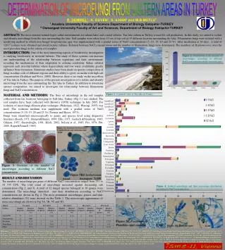
Figure 3. Decrease of the number of microfungus according to different NaCl concentration. DETERMINATION OF MICROFUNGI FROM SALTERN AREAS IN TURKEY ABSTRACT: The most extreme natural hypersaline environments are inland lakes and coastal salterns. Tuz lake saltern in Turkey is used for salt production. In this study, we aimed to isolate and identify microfungi from the area surrounding the lake. Soil samples were taken from 10 cm of top soil at 10 different location surrounding the lake. Filamentous fungi were isolated with a modifying method in which rose-bengal streptomycine agar was supplemented with a graded series of NaCl concentrations (5, 10, 15, 20 and 25 %). After incubation at 14 days, a total of 12617 isolates were obtained and stored in pure cultures. Relation between NaCl concentrations and the number of filamentous fungi were determined. The members of Hyphomycetes were the most prevalent fungi in the saltern soil samples. R. DEMIREL1, K. GUVEN1, S. ILHAN2 and M.B.MUTLU11 Anadolu University Faculty of Science Department of Biology Eskişehir-TURKEY2 Osmangazi University Faculty of Art and Sciences, Department of Biology Eskişehir-TURKEY INTRODUCTION: One of the most interesting aspects of biodiversity investigation is studying biodiversity in stressful habitats. The study of these systems can increase our understanding of the relationship between organisms and their environment, revealing the mechanisms of their adaptation to extreme conditions. Saline substrat represent such stressful habitats where hypersalinity and low water availability greatly influence biota formation.Numerous studies have been made on species composition of fungi in saline soils of different regions and their ability to grow on media with high salt concentration (Grishkan and Nevo, 2004). However, there is no study on the mycoflora of Tuz lake in Turkey. The purpose of the present investigation is to isolate and identify microfungi from the area surrounding the Tuz lake in Turkey. In addition to determine species composition, we aimed to investigate the relationship between filamentous fungi and NaCl concentration. Figure 2. Distribution of the number of microfungus according to different NaCl concentration in the research area Figure 1. A general view of Tuz lake in Turkey. MATERIAL AND METHODS: The flora of microfungi in the soil samples collected from ten locations belonging to Salt lake, Turkey (Fig 1.) was studied. The soil samples have been collected with Brown’s (1958) technique in July 2005. For isolation of microfungi dilution plate technique (Waksman, 1922; Warcup, 1955) was used. The isolation medium was supplement with a graded series of NaCl concentrations (5, 10, 15, 20 and 25 %) (Tresner and Hayes, 1971). Fungi were identified microscopically to genus and species level using diagnostic literature (Booth, 1971; Barnett&Hunter, 1999; Ellis, 1971; Gerlach &Nirenberg, 1983; Gilman, 1957; Hasenekoğlu, 1991; Klich, 2002; Nelson et al., 1983; Pitt, 1979; Pitt, 2000, Raper&Fennell, 1965). RESULT AND DISCUSSION The number of microfungi per gram of different NaCl concentration ranged from 251 to 10 735 CFU. The total count of microfungi increased against decreasing salt concentration (Fig 2. and 3).A total of 32 fungal species belonged to 10 genera were determined. The microfungi identified and their distribution according to NaCl concentration are shown in Fig 4. The most prominent microfungus genera and their relative abundance (%) were showed on the Table 1. The microscopic appearances of some microfungi are shown in Fig 5A, 5B, 5C and 5D. Figure 4. Isolated microfungi and their percentage distribution according to NaCl concentration References: 1. BARNETT, H.L., HUNTER, B.B., Illustrated Genera of Imperfect Fungi, IV. ed. 218 P. APS PRESS, USA, 1999; 2. BOOTH C, The Genus Fusarium, CAB, Kew, UK, 237 P. 1971 3.BROWN JC, Soil Fungi of Some British Sand Dunes in Relation to Soil Type and Succession. Ecology. 46: P. 641-664, 1958;4. ELLIS, M.B. Dematiaceous Hyphomycetes. CAB, Kew, UK, 608 P., 1971;5. GERLACH W, NIRENBERG H, The Genus Fusarium- a Pictorial Atlas, Biologische Bundesanstalt für Land-und Forstwirtschaft Institut für Microbiologie, Berlin-Dahlem, 406 P., 1982; 5. GILMAN JC. A Manual of Soil Fungi. The Iowa State College, Pres-Ames, Iowa, USA, 449 P., 1957;6.GRISHKAN, I., NEVO, E., Soil Microfungi of Nahal Meitsar, Golan Heights, Isreal. Plant Biosystems, 138, 21-26, 2004; 7. HASENEKOĞLU I, Toprak Mikrofungusları. Cilt I-VII, Atatürk Üni. Yay. No: 689, Kazım Karabekir Eğt. Fak. Yay. No: 11, Erzurum, 1991;8. KLICH, M.A., Identification of Common Aspergillus Species, I. ed. 122 P. Published by the Centraalbureau voor Schimmelcultures, Utrecht, The Netherlands, 2002;9.NELSON PE, TOUSSOUN TA, MARASAS WFO, Fusarium Species: An Illustrated Manual of Identification , 193 P. The Pennsylvania State University Press, University Park and London, 1983. 10. PITT, J.I., The Genus Penicillium and its telemorphic states Eupenicillium and Talaromyces, Academic Pres INC, London, 634 P., 1979; 11. PITT JI, A Laboratory Guide to Common Penicillium Species. Food Science Australia, 2000; 12. RAPER, K.B., FENNELL, D.I., The Genus Aspergillus. The Williams Wilkins Company, Baltimore, USA, 686 P., 1965; 13.TRESNER, H.D., Hayes, J.A., 1971. Sodium chloride tolerance of terrestrial fungi, Applied Microbiology, Vol. 22, No. 2, 210-21. 14. WAKSMAN SA A, Method for Counting The Number of Fungi in The Soil. J. Bacteriol. 7: P. 339-341, 1922; 15. WARCUP, J.H, On The Orgin of Colonies of Fungi Developing on Soil Dilution Plates. Trans. Brit. Mycol. Soc. 38/3: P. 298-301, 1955.