Lecture 1 Organization
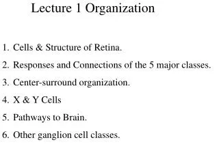
Lecture 1 Organization. Cells & Structure of Retina. Responses and Connections of the 5 major classes. Center-surround organization. X & Y Cells Pathways to Brain. Other ganglion cell classes. Retina Vertical Section. Inputs to Ganglion Cells. Classic Retinal Neuron Responses.

Lecture 1 Organization
E N D
Presentation Transcript
Lecture 1 Organization • Cells & Structure of Retina. • Responses and Connections of the 5 major classes. • Center-surround organization. • X & Y Cells • Pathways to Brain. • Other ganglion cell classes.
Early Vision & Noise • The profile of the required inhibitory field is derived from statistical estimation theory. • This profile depends strongly upon the signal:noise ratio and weakly upon the extent of lateral spatial correlation. • The receptive fields that are quantitatively predicted by the theory resemble those of X-type retinal ganglion cells and show that the inhibitory surround should become weaker and more diffuse at low intensities. • The latter property is unequivocally demonstrated in the first-order interneurons of the fly's compound eye. • The theory is extended to the time domain to account for the phasic responses of fly interneurons. • These comparisons suggest that, in the early stages of processing, the visual system is concerned primarily with coding the visual image to protect against subsequent intrinsic noise, rather than with reconstructing the scene or extracting specific features from it. • The treatment emphasizes that a neuron's dynamic range should be matched to both its receptive field and the statistical properties of the visual pattern expected within this field.
FIGURE 1. Intensity-response function of a typical visual interneuron, schematically shown here to illustrate the effect of intrinsic noise on intensity discrimination. If the response range of this spiking interneuron is restricted to, say 0-200 Hz, and if the amplitude of intrinsic noise corresponds to a fluctuation in spike frequency of, say, 10 Hz, then the neuron can encode 200/10 = 20 distinguishable intensity levels. Quantization Limits
FIGURE 2. Schematic diagram of receptive-field organization, illustrating the hypothesized encoding scheme. The signal produced by the central receptor (shown by heavy lines) is compared with a statistical prediction of this signal based on a weighted linear sum of signals from receptors in the vicinity. The interneuron encodes the difference, D, between the actual and the predicted signals, and transmits it to the higher centres. Linear Weighted Sum
FIGURE 3. The autocorrelation function of visual scenes. (a) The scene, a bed of reeds. (b) Spatial intensity profile of scene, measured along a line joining the centres of the marker discs. Intensity is plotted on a linear scale with the maximum value normalized to unity. (c) Squares: autocorrelation function of intensity profile shown in (b). Triangles: autocorrelation function of an intensity profile obtained from a horizontal scan about two disc diameters below that of (b). The autocorrelation function R(x) is computed according to R(x) = L-x I() I(6 + x) d6/ [I(6)]2d6, where I(6) is the function describing the spatial intensity profile, and L is the total length of this profile (degrees). Both x and 6 are measured in degrees. The denominator normalizes the autocorrelation function to unity, i.e. R(0) = 1. The dotted lines indicate, for each intensity profile, the ratio between the the square of the mean value and the mean square value. As x - oo the autocorrelation function should, in theory, asymptotically approach this ratio, provided that the statistical properties of the intensity fluctuations remain constant within the scene. This condition was approximately fulfilled by most of the scenes that were analysed. Autocorrelation Functions
FIGURE 5. Weighting coefficients, calculated from theory, specifying the receptive field of an encoding interneuron that receives input from an array of 11 receptors. The interneuron receives an excitatory input from the central receptor (0), and compares it with a statistical estimate of this signal derived from a suitably weighted linear combination of signals from the ten other receptors. The strengths of the inhibitory coefficients are plotted relative to that of the excitatory coefficient from the central receptor, which is normalized to unity. (The receptive-field profile of the interneuron can be derived from this array of weighting coefficients by taking into account the finite acceptance angle of each receptor.) In this and the following figures M denotes the mean intensity of the visual scene (always normalized to 1), S denotes the standard deviation of the fluctuations of intensity about the mean (i.e. the average contrast of the scene), N denotes the standard deviation of the receptor noise (expressed in terms of equivalent contrast) and D denotes the space constant of the spatial autocorrelation function of the visual scene (expressed in receptor widths). In this example the receptor signal: noise ratio (S/N) is infinity. The figure also shows the standard deviation, E, in intensity units, of the expected error between the predicted and actual values of receptor signal, calculated according to equation (1 c). Details of the calculations for this and other figures are described in Appendix 1. Weighted Surround Coefficients
Changes in Surround with Light Level (Noise) As in figure 5, except that the calculations are made for receptor signal: noise ratios of (a) IO, (b) I and (c) O.I.
Predictive Coding in Time Given that the receptor signal is temporally correlated, it should be possible to make a statistical prediction of its present value, based upon its past history. The interneuron would encode the difference between the actual present value of thereceptor signal and a statistical prediction of this value based on a weighted linear sum of past values. In this scheme, then, the role of self-inhibition would be to generate an appropriately weighted sum of the past values, and to subtract the resulting prediction from the excitatory signal corresponding to the present value. FIGURE 9. Predictive coding in the time domain. The figure shows temporal weighting functions calculated for receptor signal:noise ratios of (a) 10, (b) 1 and (c) 0.1. Time is depicted in units of receptor integration time, and the parameter D now refers to the time constant of the temporal autocorrelation function of the receptor signal, specified in these units. All other parameters have the same meaning as in earlier figures.
Center & Surround Responses: Suppressive vs. Antagonistic Surrounds