Download
1 / 50
660 likes | 1.03k Vues
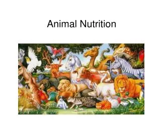
ANIMAL NUTRITION. Diverse Feeding Adaptations. Suspension Feeders – sift food particles from water. Substrate Feeders – eat their way through their food source. Deposit Feeders – eating their way through partially decayed matter. Fluid Feeders – suck fluids out of a living host.

E N D
Diverse Feeding Adaptations • Suspension Feeders – sift food particles from water. • Substrate Feeders – eat their way through their food source. • Deposit Feeders – eating their way through partially decayed matter. • Fluid Feeders – suck fluids out of a living host. • Bulk Feeders – eat large pieces of their prey.
Four Main Stages of Food Processing • Ingestion- act of eating. • Digestion – process of breaking food down into molecules that can be absorbed by the body. • Absorption – absorbing the nutrients into the blood. • Elimination – ridding the body of waste.
Digestion Occurs in Specialized Compartments • Prevents enzymatic hydrolysis of the organisms own tissues. • Food vacuoles. • Gastro vascular cavities. • Alimentary cavities.
Intracellular Digestion Verses Extra Cellular Digestion. • Intracellular Digestion • Food vacuoles ingest food particles that fuse to a lysosome containing hydrolytic enzymes. • Heterotrophic protists and sponges do this. • Extracellular Digestion • Occurs outside of cells. • Gastrovascular cavities – hydra, planaria • Incomplete digestion – mouth and anus are the same opening • Digestive enzymes secreted by the gastrodermal cells digest the prey into tiny bits.
Alimentary Cavities. • Digestive tube • Complete digestive tract – mouth and anus at opposite ends. • Nematodes, Annelids, Mollusks, Arthropods, Echinoderms and Chordates. • Enzymes are secreted by the digestive tube and nutrients are absorbed along the way.
Specialized Structures of the Digestive Tube • Mouth and pharnyx – where food is taken in. • Esophogus - passage • Crop – food moistened and stored (insects and birds and annelids) • Gizzard – food is pulverized (birds and annelids) • Intestine – enzymes digest food and nutrients absorbed. • Anus – where undigested food and waste exits.
The Mammalian Digestive System • Peristalsis is the rhythmic contractions of smooth muscle along the digestive tract that keep food moving along. • Sphintors close off various parts of the tube so that regulating the passage of food between chambers. • Salivary Glands secrete amylase to break down starch and lubricate food. • Pancrease secretes digestive enzymes. • Liver produces bile. • Gallbladder stores bile.
The Oral Cavity • Chewing physically breaking the food into smaller pieces and exposing more surface area for enzymes to act on. • Salivary amylase breaks down starch. Aliva also contains mucin with makes food slippery. • Bolus is the ball of food that gets pushed into the esophogus.
The Stomach • Can accommodate about 2 liters of food. • Chyme is what the bolus becomes in the stomach after digestive juices are added. • Ridges in the stomach called ruggae help churn food. • Pepsin breaks down proteins. • Pepsinogen is the inactive for of pepsin and is activated by HCl. • HCl also helps denature proteins in the chyme.
Digestion in the Stomach A Closer Look • Gastric Pits in the stomach lining are composed of specialized epithelial cells. • Gastric pits create the gastric juices. • Parietal Cells secrete HCl. • Chief Cells secrete pepsinogen. • Mucus Cells secrete mucus that protects the stomach lining.
Feedback Systems • Pepsin activates more pepsinogen. • The sight of food creates a nervous response that causes gastric juices to be secreted. • A drop in pH and distention of the stomach creates a negative feedback response.
Problems • Gastric ulcers are lesions in the stomach lining. • Can occur when the lining is eroded faster that it is replaced. • Helibacter pylori can cause ulcers but can be treated with antibiotics. • Heartburn occurs when acid chyme seeps back through the cardiac sphintor which is the opening between the esophogus and stomach.
Enter the Small Intestine! • The pyloric sphinctor is the opening between the small intestine and the stomach. • It takes 2 to 6 hours after a meal for the stomach to empty. • The small intestine is known as the duodenum. • The small intestine secretes bicarbonate to neutralize the acid chyme coming from the stomach. • Bile is secreted by the gallbladder to emulsify fats which are broken down by an enzyme called lipase which is secreted by the small intestine.
Enzyme Digestion • Carbohydrates • Pancreatic amylasesecreted by the pancreas breaks down starch. • Maltase, Sucrase, Lactase break down disacharrides and are built into the intestinal epithelium. • Protein Disgestion • Chymotrypsin, Trypsin,Carboxypeptidase, Aminopeptidase, enteropeptidase. • Nucleic Acids – Nucleases • Lipids - Lipases
Absorption of Nutrients • Villi and microvilli increase surface area(300m2) for maximum absorption. • Intestinal epitheium absorb nutrients either by diffusion or active transport. • Nutrients are carried away from the intestine by capillaries at the core of the villi. • Lactealsare lymphatic vessels that are surrounded by capillaries in the core oif the villie that absorb fats which are combined with proteins. • These lipid proteins are calledchylomicrons. • They travel from through the lyphatic system and eventually drain back into the blood and travel to the heart.
Capillaries and veins drain blood into the hepatic portal vessel which carries blood to the liver. • Ensures that the liver has first access to the nutrients in the blood. • The nutrient balance of the blood leaving the liver may be very different than it was when it entered. • One of the many functions the liver include regulating glucose levels in the blood and converting amino acids into carbohydrates. • From the liver the blood travels to the heart to be pumped to the rest of the body.
Hormonal Regulation of Digestion Hormones released by the stomach and duodenal wall ensure that digestive juices are only around when they are needed. The sight of food will stimulate the brain to tell the stomach wall to release gastrin which in turn stimulates gastric juices to be secreted. Gastric juices cause more gastric juices to be released. A drop in pH inhibits gastrin.
Enterogastronesare secreted by the duodenal wall which causes the duodenal wall to secrete secretinwhichsignals the pancreas to secrete bicarbonate to nueteralize chyme as it enters the duodenum. • Cholecystokinin(CCK)siganls the gallbladder to contract and secrete bile into the duodenum when chyme is rich in fats. • Other enterogasterones restrict peristalsis when chyme is rich in fats.
Reabsorbing Water • Most of the water from waste is absorbed in the colon. • The junction between the duodenum and the colon is called the Cecum. • Humans have a small ceum compared to other animals. • A fingerlike projection that extends from the cecum is called the appendix.
Colon Bacteria • Flora are bacteria that live within the body that are beneficical. • E.Coli live in the colon and produce vitamins, K B, biotin and folic acid. • Generate gases such as methane and hydrogen sulfide as by products of microbial metabolism.
Structual Adaptations of the Digestive System • The type of teeth and the way that they are arranged accommodate an animals’s diet. • Herbivores like horses have flat molars fro grinding. • Carnivores have pointed incissors for tearing and catching prey. • Carnivores have large expandable stomachs to allow them to eat large a mounts of food at once because there may be long periods of time where they go without food. • Snakes have hollow teeth to store venom. • Snakes have a flexible ligament that allows them to open their jaw to swallow prey whole.
Length of Digestive Tract • Herbivores and Ominvores have a relatively long alimentary cavity relative to their digestive tract because they ingest cellulose. • Hard to digest and low in energy content. • Many herbivores have symbiotic bacteria living in specialized organs that digest cellulose • Ruminants • Crop in the Hoatzin(South American Bird from the Rain Forest) have bacteria that digest cellulose. • Rabbits eat their feces and digest it again. • Cows regurgitate and chew their food over again.