Download
1 / 22
220 likes | 576 Vues
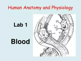
Human Anatomy and Physiology. Lab 1 Blood. Background: I. Blood is a connective tissue composed of formed elements (cells and cell fragments) and intercellular material (plasma). Blood Sample Centrifuged. Objective 1 Plasma Characteristics

E N D
Human Anatomy and Physiology Lab 1 Blood
Background: I. Blood is a connective tissue composed of formed elements (cells and cell fragments) and intercellular material (plasma)
Objective 1 Plasma Characteristics Plasma is the extracellular (intercellular) material of the connective tissue, blood: Characteristics: 90% H2O 10% solutes: plasma proteins (albumin, antibodies, clotting proteins) nutrients (eg, proteins, carbohydrates, lipids, vitamins) hormones wastes (eg, urea, uric acid, creatinine) dissolved gases (CO2, O2)
Lab Objective 1: In this exercise, you will obtain a sample of sheep plasma and determine its: color clarity pH consistency
Formed Elements: cells and cell fragments Red Blood Cells (RBCs): anucleate cells that carry oxygen; there are 4-6 X 10⁶/mm3 blood White Blood Cells (WBCs): nucleated cells that provide immunity; there are 5,000 – 9,000/mm3 blood Platelets (thrombocyte) cell fragments used for hemostasis (stoppage of bleeding); there are 250,000 – 400,000/mm3 blood
Red Blood Cells (erythrocytes): • 1. Are biconcave discs that lack nuclei and organelles • 2. Contain hemoglobin (Hb) which binds to O2 • 3. They are the most numerous formed element • 4. They have a “biconcave” shape • 5. Their diameter averages 7.5 m.
There are five types of leukocytes organized into two classes: Agranulocytes: Granulocytes: lymphocytes basophils monocytes neutrophils eosinophils
Platelets (Thrombocytes): • 1. Are fragments derived from bone marrow cells (megakaryocytes) • 2. They contain granules but no nuclei or organelles • They are smaller than RBCs • They are used to prevent blood loss (hemostasis)
Objective 2 You will identify each of the formed elements on a prepared blood smear that has been stained with Wright’s stain Wright’s Stain is a mixture of two dyes: 1. Methylene Blue: a basic dye that stains acidic components deep blue/purple 2. Eosin: an acidic dye that stains basic components red/deep pink/orange
Different stains can be applied – one popular stain is a differential stain called Wright’s Stain. Wright’s stain is a mixture of eosin and methylene blue. • Methylene blue (blue dye) • has a positive charge and stains negatively charged substances (acids found in some granules, and DNA and RNA) • structures that combine with methylene blue are called basophilic Eosin (red dye) has a negative charge and stains positively charged substances (bases found in some granules, and hemoglobin) structures that combine with eosin blue are called acidophilic Eosin/Methylene Blue Complex stains neutral substances (such as components of some granules) lilac
When blood is smeared onto a slide, dried and stained with Wright’s stain, the individual formed elements (including types of WBC’s) can be distinguished: Erythrocyte Leukocyte Platelet
Granulocytes: Neutrophil: 40 – 70% of the circulating WBCs - 9-16 m in diameter - 2-5 nuclear lobes - pale staining, lavendar granules
Eosinophils: - 1-4% of the total circulating WBCs - 10-14 m in diameter - have a bilobed nucleus - bright reddish/orange/pink cytoplasmic granules
Basophils: - 0 – 1% of the total circulating WBCs - 8-10 m in diameter - unsegmented or biloed (usually) nucleus - deep blue/purple cytoplasmic granules
Agranulocytes: Lymphocyte: - 20 - 45 % of the total WBC count - size ranges from 5 m (small) to 17 m (large); small lymphocytes predominate - nucleus is round or slightly oval; it may be indented - clear blue cytoplasm that may be seen only as a ring around the nucleus Small lymphocyte Large lymphocyte
Monocyte - 4-8 % of the total WBC count - 14-24 m in diameter - nucleus is horseshoe shaped or kidney shaped - abundant blue gray cytoplasm that may contain vacuoles
Platelets appear as small, granular cell fragments (2-3 m in diameter) that may occur singly or in clumps
Objective 3 A differential white blood cell countis performed to determine the relative percentage of each type of WBC It is used to detect diseases, such as acute infection, chronic infection, allergy, parasitic diseases, anemia, HIV infection, and others
In this activity, you will systematically scan a prepared slide and observe 100 white blood cells - identify each one and record your data to determine the relative percentages of each type of WBC # observed %= X 100 # counted
Each of the leukocytes exists within a range of normal values in peripheral blood. Type of LeukocyteNormal %#/100 CellsIf Elevated? Neutrophil 40-70% 40-70 acute infection Eosinophil 1-4% 1-4 allergic reaction, parasitic infection Basophil <1% 0-1 ??? Lymphocyte 20-45% 20-45 Monocyte 4-8% 4-8 chronic infection