Download
1 / 15
150 likes | 170 Vues
Explore autonomic pathways in the head through animations with key landmarks and a self-testing feature. Educational module by Bob Hutchins, PhD, from Baylor College of Dentistry.

E N D
Autonomic Innervation to the Head Animations and a Self-Testing guide Bob Hutchins, PhD, Dept. of Biomedical Sciences, Baylor College of Dentistry, Texas A&M University System HSC 2008
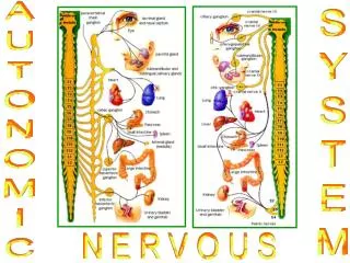
Contents • This module illustrating the Autonomics to the head is presented in 3 parts: • Part I provides an overview of the educational objectives • Part II provides an animated view of each of the four parasympathetic pathways as well as the sympathetics to the head • Part III is a repeat of the same animated views as in part II, however each of the views have important landmarks numbered with an accompanying key • *This last part has been designed as a self-testing feature Jump to Part I Jump to Part II Jump to Part III Bob Hutchins, PhD, Dept. of Biomedical Sciences, Baylor College of Dentistry, Texas A&M University System HSC 2008
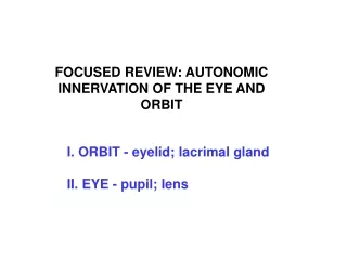
Part I - Educational Objectives • It is expected that once you have finished this module, you will be able to: • Identify the preganglionic parasympathetic autonomic pathways to the head from their nuclear origin to their point of termination • Identify the postganglionic parasympathetic autonomic pathways to the head from their ganglionic origin to their termination in the effector organ • 3. Recognize how the postganglionic sympathetic neurons travel to their site of termination in the effector organ • 4. Be able to identify important landmarks associated with the autonomic pathways in the head. Bob Hutchins, PhD, Dept. of Biomedical Sciences, Baylor College of Dentistry, Texas A&M University System HSC 2008
Part II • The following are animated slides demonstrating the autonomic pathways to the head. Bob Hutchins, PhD, Dept. of Biomedical Sciences, Baylor College of Dentistry, Texas A&M University System HSC 2008
Preganglionic parasympathetics Postganglionic parasympathetics Postganglionic sympathetics Ciliary Ganglion
Preganglionic parasympathetics Postganglionic parasympathetics A B Submandibular Ganglion
Preganglionic parasympathetics Postganglionic parasympathetics Postganglionic sympathetics Pterygopalatine Ganglion
Preganglionic parasympathetics Postganglionic parasympathetics Postganglionic sympathetics Otic Ganglion
Part III • The following slides are the same drawings to the head with areas along the pathway numbered. An accompanying key (either on the same slide or on the following slide) is included to assist you in reviewing the bony landmarks and learning the typical autonomic pathways to the head.
1. Edinger-Westphal n. • 2. Oculomotor n. (CN III) • 3. Superior orbital fissure • 4.* Ciliary ganglion • 5. Short ciliary nerves • 6. Eye • 7. Nasociliary nerve • 8. Long ciliary nerves • 9. Internal carotid plexus • 10. Internal carotid a. • 11. Optic n. (CN II) • Optic canal • 20. Trigeminal ganglion • 21. Mandibular division of CN V (CN V3) • 22. Maxillary division of CN V (CN V2) • 23. Ophthalmic division of CN V (CN V1) • 24. Maxillary sinus Ciliary Ganglion • Cross-section of the brain stem illustrating the nuclear origin of the parasympathetic fibers • B. Figure illustrating peripheral distribution of the parasympathetics and the sympathetics
1. Superior salivatory nucleus • Facial n (CN VII) • (nervus intermedius) • 3. Internal auditory meatus • 4. Geniculate ganglion • 5. Malleus • 6. Incus • 7. Petrotympanic fissure • 8. Chorda tympani • 9. Lingual n. (mandibular div. of CN V) • 10.* Submandibular ganglion • 11. Submandibular gland • Lingual gland • 20. Trigeminal ganglion • 21. Foramen ovale • 22. Stylomastoid foramen • 23. Facial n. (CN VII) • 24. Tongue Submandibular Ganglion • Note: The sympathetics from the superior cervical ganglion travel with the appropriate • arteries to this area of the face via the external carotid a. • Cross-section of the brain stem illustrating the nuclear origin of the parasympathetic fibers. • B. Figure illustrating the peripheral distribution of the parasympathetics.
Pterygopalatine Ganglion NOTE, the numbered key for the autonomic landmarks are found on the next slide.
Pterygopalatine Ganglion 1. Superior salivatory nucleus 2. Facial n (CN VII) 3. Internal auditory meatus 4. Geniculate ganglion 5. Greater petrosal n. 6. Hiatus of the facial canal 7. Deep petrosal n. 8. Foramen lacerum 9. Nerve to the pterygoid canal 10. Lesser and greater palatine nn. 11.* Pterygopalatine ganglion 12. Nasopalatine nerve 13. Zygomatic nerve 14. Zygomaticotemporal nerve 15. Lacrimal nerve 16. Lacrimal gland 17. Nasopalatine nerve 18. Incisive foramen 19. Lesser palatine nerve 20. Greater palatine nerve 21. Facial nerve (CN VII) 22. Internal jugular v. 23. Internal carotid a. 24. Internal carotid plexus 25. Carotid canal 26. Trigeminal ganglion 27. Sella turcica 28. Internal carotid a. 29. Ophthalmic division of CN V (CN V1) 30. Maxillary division of CN V (CN V2) passing through the foramen rotundum 31. Posterior superior alveolar n. 32. Infraorbital nerve 33. Inferior orbital fissure 34. Zygomaticofacial nerve 35. Inferior alveolar nerve 36. Stylomastoid foramen 37. Zygomatic bone 38. Chorda tympani 39. Semi-circular canals 40. Choclea. 41. Sphenopalatine foramen • Cross-section of the brain stem illustrating the nuclear origin of the parasympathetic fibers • Figure illustrating the peripheral distribution of the parasympathetics and the sympathetics • C. Figure illustrating the continuation of the nasopalatine nerve as it passes from the spehenopalatine foramen laterally onto the midline nasal septum Also note numbers 19 and 20 which represent the terminal position of these nerves onto the palate minor salivary glands.
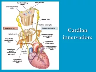
Inferior salivatory nucleus Glossopharyngeal n. (CN IX) Jugular foramen Internal jugular v. Glossopharyngeal n. (CN IX) Internal carotid a. Internal carotid plexus Tympanic plexus Lesser petrosal n. Hiatus for the lesser petrosal n. Trigeminal ganglion Foramen ovale *Otic ganglion Mandibular division of CN V (CN V3) Auriculotemporal n. Parotid gland Internal auditory meatus Stylomastoid foramen Middle ear cavity Ophthalmic division of CN V (CN V1) Maxillary division of CN V (CN V2) Otic Ganglion • Cross-section of the brain stem illustrating the nuclear origin of the parasympathetic fibers. • B. Figure illustrating the peripheral distribution of the parasympathetics and the sympathetics. **I would like to credit Baylor’s media department in their assisting me in the drawing of the skull material.
Inferior View of the Skull Demonstrating the Exit of 3 Parasympathetic Nerves 2 3 1 4 • The greater petrosal n.(CN VII) begins to descend through the F. Lacerum, joins with the deep petrosal n. (sympathetics) and before exiting the F. Lacerum, enters the pterygoid canal anteriorly (arrow head). • CN V3 exiting the F. Ovale. • The auriculotemporal n. branching from CN V3 and carrying with it the postgang. parasympathetic nerves traveling to the parotid gland. • The chorda tympani exiting the petrotympanic fissure and carrying with it the pregang. parasympthetic nerves to the submandibular gland.