WHAT IS THE DIAGNOSIS
490 likes | 694 Vues
WHAT IS THE DIAGNOSIS. Prof. Dr. İnci GÜLMEZ Erciyes University Medical Faculty Department of Pulmonology, Kayseri. Case. 36 years old, woman with two children housewife, hometown Pınarbası, lives in Kayseri. 2004. Cough , exercitional dyspnea

WHAT IS THE DIAGNOSIS
E N D
Presentation Transcript
WHAT IS THE DIAGNOSIS Prof. Dr. İnci GÜLMEZ Erciyes University Medical Faculty Department of Pulmonology, Kayseri
Case 36 years old, woman with two children housewife, hometown Pınarbası, lives in Kayseri
2004 • Cough , exercitional dyspnea • Diagnosed with asthma five years ago • Four years history of joint pains, finger swelling and elevated RF, diagnosed wtih rheumatoid arthiritis • Medications: NSAIDs, Leflunomide (Arava) • Her mother is affected with asthma • Chest X-ray: bilateral reticulonodular infiltration and apical consolidation
2004 • Physical examination: has a BCG scar Long expiratory time , bilateral ronchus and ralles • Laboratory: Hb:11,8gr/dl WBC:5300/mm3 Sedimentation: 52mm/h RF:276Ü/mL( 0-20) CRP:215 mg/L(0-200)
What is the differantial diagnosis? • Tuberculosis • Rheumatoid lung • Drug induced pulmonary disease • Caplan’s syndrome
2004 • Spirometry: FVC %70 FEV1 %48 FEV1/FVC: %60 • DLCo:%82 • ADA:15Ü/ml( cut of point 25 U/ml) • ANA(+) • Sputum EZN: Three times (-) • Bactec and L-J culture: negative • PPD: 7 mm/ 72 h Watts RA et al Semin Arthritis Rheum.1995;25:28–34
2004 • Bronchoscopy : BAL EZN(-) • BAL Bactec and L-J: negative • BAL cytology: Clas II • Bronchoscopic Bx: no lung tissue, normal bronchial tissue • ECG: normal • ECHO: EF: %68, PAP: 25 mmHg
What is the diagnosis? • Tuberculosis • Rheumatoid lung • Drug induced pulmonary disorders
What is the diagnosis? • Tuberculosis • Rheumatoid lung • Drug induced pulmonary disorders
THERAPY • Oral steroid (for one year) • ICS+LABA • NSAIDs
June 2006 • Had been taking NSAID, ICS +LABA for eight months • Joint pain • Physical examination: additional sign: small joints of the hands are inflamed • Laboratory: Hb: 11,9gr/dl Sd: 32mm/h RF:606 and 1070 U/mL • ANA:+ CRP:79,3 mg/L
June 2006 • Spirometry : Baseline: FVC%87 FEV1:%63 FEV1/FVC: %63 Post bronchodilatör: FVC:%88 FEV1:%66 FEV1/FVC:65 • DLCo: %74
Therapy • Oral steroid • ICS+ LABA • NSAİDs
June 2007 • 10 mg florokortolon, ICS+LABA, NSAID • Exercitional dyspnea and chest pain • Exercitional palpitation • ECG: Right axis, right ventricular hypertrophy
What test would you choose at first at this step? • HRCT • RF • DLCo • ECHO • Spirometry • 6 MWT
What test would you choose at first at this step? • HRCT • RF • DLCo • ECHO • Spirometry • 6 MWT
June 2007 • ECHO: 2nd degree T sufficiency, • PAP: 60-65 mmHg, • Left ventricular function is normal
June 2007 • RF: 69 U/mL • DLCo: %72 • Spirometry: postbronchodilatory FVC: %66 FEV1.%46 FEV1/FVC: 60 • 6 MWT: 310 M, heart rate at the end of the test :145/min
What is the additional suggestion in therapy? • Teophylin • Spironolactone • Bosentan • İlomedin • Calcium canal blocker • Sildenafil
What is the additional suggestion in therapy? • Teophylin • Spironolactone • Bosentan • İlomedin • Calcium canal blocker • Sildenafil
One month later • Complaint of dyspnea • ABG ( room air):PH: 7,52 PaCO2:29mmHg PaO2:79 mmHg • DLCo: %52 • Spirometry: FVC:%51 FEV1: %40 FEV1/FVC: %68 • 6MWT: 270 M
Therapy • Prednizolon 60mg+ cyclophospamide 100mg • Diltiazem 3x60 mg • ICS+ LABA • Anticoagulant
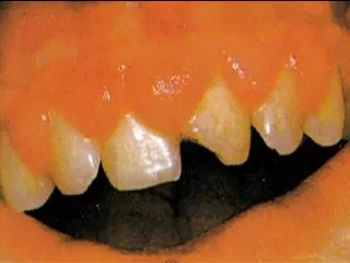
October 2007 • Oral lesions • Elevation in dyspnea symtoms • Palpitations • Sweating • Chest pain • Lab: WC: 25300/mm3 sed:96 mm/h • Hospitalisation
Loss of volume on the right hemithorax • Increase in the pulmonary artery diameter • Increase of parietal pleural nodules on right hemithorax • Mass in the anterior mediastinum • Increase of ascendan aorta diameter
Loss of volume on the right hemithorax • Increase in the pulmonary artery diameter • Increase of parietal pleural nodules on right hemithorax • Mass in the anterior mediastinum • Increase of ascendan aorta diameter
October 2007 • Kandidomannan: positive • freeT3-freeT4- TSH: normal • Sputum gram stain: PK(+), no microorganism is seen • Galaktomannan: 0,27 (-) • CRP: 184mg/L
What do you think about differential diagnosis? • Tuberculosis • Candida pneumonia • İnvazive aspergillosis • Cyclophospamide toxicity • Thymoma • Actinomycosis • Nocardiosis
Additional Therapy • Liposomal Amphotericin B 1mg/kg • Moxifloxasin 400mg/day
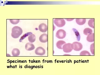
Oral culture : (-) • Blood culture: (-) • Suputum microscopy: no fungus is found • Sputum culture: candida albicans • Suputum EZN: negative • Bronchoscopy: enflamation , BAL is taken from upper left lobe, and TBBx is taken from apicoanterior upper left lobe
Third day • Reduced general contidition • Tachycardia unresponsive to medical treatment • Hypoxaemia despite Oxygen therapy mask use • Control X-ray
Control Chest X-Ray Intralesionar punction: pus
Third day • Pus material EZN stain: negative Gram stain: PK(+), suspicious gram pozitive microorganism • Lavage culture: No reproduction • Patient’s Hypoxaemia went deeper, respiratory failure, mechanic ventilation support, exitus!
What is the diagnosis? • Tuberculosis • Necrotizing pnomonia (resistant microorganism) • İnvasive aspergillosis • Actinomycosis • Nocardiosis
Pus material : Nocardia spc Specification is not done
Nocardiosis • Nocardiosis is an acute, subacute, or chronic suppurative infection caused by Nocardia • Weakly gram-positive, filamentous bacteria found worldwide in soils • A total of approximately 30 strains of Nocardia have been associated with human disease • Infections are localized or dissemineted
Nocardiosis • Disseminated and fulminant disease mainly occurs in immunocompromised host with underlying illnesses, such as HIV, cytotoxic chemotherapy, bone marrow transplantation, or prolonged glucocorticoid treatment
NocardiosisClinical manifestations • Bronchopneumonia • Lobar pneumonia • Necrotizing pneumonia • Mediastinitis • Mediastinal abscesses • Cerebral abscesses • Intra-abdominal abscesses • Peritonitis
Nocardiosis • The primary disease occurs in the pulmonary system and may mimic tuberculosis, staphylococcal, or mycotic infections • Hematogenous dissemination may occur to all organs of the body • The brain, kidneys, and liver are the most common metastatic sites
Nocardiosis • Mortality is increased in patients with acute infection and in those with disseminated disease involving two or more contiguous organs or the CNS • Mortality is also increased in patients taking corticosteroids or antineoplastic agents
NocardiosisPhysical examinations • Subacute abscesses are palpabl at the site of trauma and generally fell firmer than fluctuant • A lung examination may reveal diffuse or localized abnormal breath sounds • Mild-to-severe respiratory distress that progresses to respiratory failure may occur
Nocardiosisİmaging studies • Generalized infections - chest radiographic findings vary and include fluffy infiltrates, scattered nodules, and confluent lobar infiltrates progressing to complete consolidation and cavitation - Chest CT scanning may be necessary to visualize the extent of disease and to rule out empyema
NocardiosisDiagnosis • Gram-stain -Directly examine clinical materials (eg, sputum, bronchoalveolar lavage, cerebral spinal fluid, pus) by gram stains and acid-fast stains -Use methamine-silver stains for demonstrating the organisms in tissue specimens • Serological diagnosis is not readily available • Cultures typically grow within 3-5 days on blood or chocolate agar
NocardiosisTreatment • Sulfa-based therapy is recomended. TM-SM, given intravenously in high doses, is the treatment of cohoice • Linezolid has a growing literature in support of its use in combination and monotherapy • Additional concurrent therapy with an aminoglycoside plus ceftriaxone benefits patients with fulminant disease
NocardiosisTreatment • Patients who are immunocompetent with lymphocutaneous disease are usually treated for 6-12 weeks • Therapy includes incisions and drainage of abscesses • Patients with immunocompromising conditions are treated for at least three months after clinical cure( usually up to one year of threapy)