How to read chest x-ray
250 likes | 442 Vues
How to read chest x-ray. Dr. Suheab A. Maghrabi MBBS, MSc. X rays: Electromagnetic R adiation. Absorption varies between tissues. Bone Soft tissue Air . Be systematic. Don’t rush to conclusions. P atient profile. Name. Date. T ype of view. Image Quality: Penetration

How to read chest x-ray
E N D
Presentation Transcript
How to read chest x-ray Dr. Suheab A. Maghrabi MBBS, MSc.
X rays: Electromagnetic Radiation. • Absorption varies between tissues. • Bone • Soft tissue • Air
Be systematic. • Don’t rush to conclusions.
Patient profile. • Name. • Date. • Type of view. • Image Quality: • Penetration • Inspiration • Rotation • Angulation • Soft tissues / bony structures • Mediastinum • Diaphragms • Lung Fields
Patient Profile • Name. • Date.
Image Quality: • Penetration. • Inspiration. • Rotation. • Angulation.
Rotation: • Distance between clavicle and spines process should be equal on both sides.
Angulation: • Clavicle should lie on the 3rd rib. 3
Soft tissues / bony structures • Symmetry • Deformities • Fractures • Masses • Calcifications • Lytic lesions
Mediastinum • Heart • Shape. • Sharpe edges. • Enlargement. • Hilar Lymph nodes and vessels.
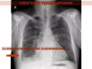
Diaphragms • free air. • Gastric buble (left) • Margins should be sharp. • Right dome usually higher than left. • Clear Costophrenic angle.
Lung Fields • Infiltrates. • Increased interstitial markings. • Masses. • Air bronchograms. • Increased vascularity. • Lobs and fissures.