FOCUS
FOCUS. Foundation - firm Observation – landmarks Concentration – in the moment, mindfulness Understanding – data, information, knowledge, mastery, wisdom Systematic – Thought process of expert. The Warrior & the Surgeon 13 July 2009. Dr. Frank C.T. Voon antvoon@nus.edu.sg.

FOCUS
E N D
Presentation Transcript
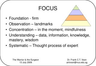
FOCUS • Foundation - firm • Observation – landmarks • Concentration – in the moment, mindfulness • Understanding – data, information, knowledge, mastery, wisdom • Systematic – Thought process of expert The Warrior & the Surgeon 13 July 2009 Dr. Frank C.T. Voon antvoon@nus.edu.sg
Medical Diagnosis Symptoms Signs Investigations
Medical Education Signs Symptoms Structures Systems Regional Anatomy Functional Anatomy Specialties Imaging Anatomy
Structures • Bones • Joints • Muscles • Nerves • Arteries • Veins • Lymphatics • Organs • Structures • Compartments
Systems • Skeletal • Articular • Muscular • Nervous • Endocrine • Circulatory – cardiovascular • Immune - lymphatic • Respiratory • Digestive • Excretory - renal • Reproductive – female & male • Integumentary
The Analytically Anatomical Approach • Recognize the region(6) & subregion • Determine the perspective • Anterior, posterior, sagittal, superior, inferior • Locate the level • Superficial (skin) to deep (bones) • Vertical levels (vertebra or plane) • Select the structure • B J M N A V L O S C • Choose the format
Regions • Head & neck • Upper limb • Thorax • Abdomen • Pelvis • Lower limb
Region - Head • Face • Scalp • Temporal region • Infratemporal region • Orbit • Nasal cavity • Oral cavity • Ear - outer
Region - Neck • Suprahyoid, infrahyoid • Triangles – anterior & posterior • Pharynx • Larynx • Carotid sheath
Region - Upper limb • Shoulder • pectoral, scapular • Arm • Flexor and extensor compartments • Forearm • Flexor and extensor compartments • Hand • Thenar, intermediate, hypothenar compartments • The free upper limb, pectoral girdle
Region - Lower limb • Gluteal region • Thigh • Anterior compartment - extensor • Medial compartment - adductor • Posterior compartment - flexor • Leg • Anterior compartment - extensor • Lateral compartment - peroneal • Posterior compartment - flexor • Foot • Layers of the foot • Muscle • Tendon • Muscle • Bone
Region - Thorax • Boundaries • Walls • Anterior – manubrium, sternum, xiphisternum, costal cartilages • Lateral – ribs, intercostal spaces • Posterior – vertebrae, intervertebral discs, erector spinae • Roof – thoracic inlet/outlet • Floor – thoracic outlet/diaphragm • Contents • Pleural cavities • Mediastinum • Superior • Trachea, arch of aorta, esophagus, superior vena cava, brachiocepalic trunk, left common carotid artery, left subclavian artery • Inferior • Anterior – thymus, internal thoracic vessels • Middle – pericardium, heart, pulmonary trunk, ascending aorta • Posterior – esophagus, descending aorta, azygos system, thoracic duct
Region - Abdomen • Boundaries • Anterolateral abdominal wall • rectus abdominis • external oblique, internal oblique, transversus abdominis • Posterior abdominal wall • Psoas major, quadratus lumborum • Inguinal region • Contents • Compartments • Peritoneal cavity • Organs • Gut – foregut, midgut, hindgut • Liver, pancreas, spleen • Kidneys
Region - Pelvis • Boundaries • Anterolateral abdominal wall • rectus abdominis • external oblique, internal oblique, transversus abdominis • Posterior abdominal wall • Psoas major, quadratus lumborum • Inguinal region • Contents • Compartments • Pouch of Douglas, rectovesical pouch, uterovesical pouch • Organs • Urinary bladder, prostate, seminal vesicles • Uterus, vagina • Rectum • Perineum • Anal triangle, urogenital triangle, urogenital diaphragm
Bones • Type • Long • short • flat • irregular • sesamoid • Upper end, lower end, shaft • Projections – tuberosities, condyles, neck • Right or left – all 3 dimensions • Ossification – endochondral, intramembranous
Bones • Mandible – temporomandibular joint, parts • Vertebrae – cervical, thoracic, lumbar, sacrum • Rib – typical, 1st • Clavicle – intramembranous, sternoclavicular joint • Scapula – muscles, rotation 60 degrees • Humerus – surgical neck, midshaft, supratrochlear, medial epicondyle • Radius – pivot joint, tuberosity (biceps) • Carpus – scaphoid, trapezium, capitate, hamate • Femur – neck, head, lesser trochanter (iliopsoas) • Fibula – lateral malleolus (key to orientation) • Patella – sesamoid bone, larger lateral facet • Tarsus – talus (ball bearing)
Joints • Definition • Articulation between 2 or more bones • From outside in • Capsule • Synovial membrane • Articular cartilage • Ligaments • Movements allowed
Joints • TMJ - dislocation • Atlanto-occipital – flexion/extension & lateral flexion, condyloid joint • Atlanto-axial - pivot joint • Intervertebral – cartilaginous joint • Sternoclavicular – double plane • Thoracic cage – bucket handle, pump handle • Shoulder, wrist, elbow – ball & socket, ellipsoid, hinge • Hip, knee, ankle • 1st carpometacarpal • Axis of movements • Supination, pronation, inversion, eversion
Muscles • Origin – bone, then part • Insertion – bone, then part • Joint/s involved • Movements possible – use the joint axis • Nerve supply • Blood supply – arteries, veins, anastomosis • Relations - • Applied/clinical anatomy – major function,
Muscles – H&N • Muscles of mastication – 1st arch • Muscles of facial expression – 2nd arch • Extra-ocular muscles – LR6,SO4 • Soft palate • Tongue • Supra- & infra- hyoid muscles • Sternocleidomastoid & trapezius • Scalenus anterior & medius
Muscles – UL • Upper – shoulder & arm • Scapular group • Triceps • Flexors • Coracoid process • Pectoral girdle & humeral • Lower – forearm & hand • Flexors – superficial & deep • Extensors – lateral, thumb, digits, elbow • Thenar, intermediate, hypothenar
Muscles – LL • Thigh – extensors, adductors, flexors • Gluteal & lateral rotators • Leg - extensors, peronei, flexors • Foot – muscle, tendon, muscle, bone
Muscles – Trunk • Intercostals • Diaphragm • Obliques & transversus abdominis • Pelvic diaphragm • Urogenital diaphragm • Erector spinae
Muscles – smooth & cardiac • Intrinsic muscles of the eye • Stapedius & tensor tympani • Musculi pectinati, trabeculae carneae, • Detrusor • Gut – colic, outer longitudinal, inner circular • Oesophagus – skeletal to smooth, 1/3 • Sphincters of urethra & anal canal
Nerves • Origin • Root levels • Branches • Distribution • Clinical - palsies
Nerves • Cranial - trigeminal, facial & vagus • Spinal • ARMUM • FOGS • Tibial; common, superficial & deep peroneal • Plantar – medial & lateral • Autonomic • COPS • Splanchnics - greater, lesser & least; pelvic
Arteries • Begins • A continuation of • Location, usually bony part nearby • Ends (as above) • Middle • If 3 parts, middle related to a muscle • 4 parts (vertebral artery) • branches • distribution • Relationships • V-A-N • Histological layers • Endothelium, muscle (smooth), adventitia (elastic) • Applied • Anastomosis, clinical procedures
Arteries • External carotid • Soft palm • ST,L,F,O,PA,AP,ST,Mx • Internal carotid - circle of Willis • Aorta – ascending, arch, descending, abdominal • Internal iliac • Femoral, tibial, popliteal, plantar • Brachiocephalic, subclavian, axillary, brachial, radial, ulnar, palmar arches (superficial & deep) • Coronary arteries • Choroid plexuses, anastomoses, coarctation of the aorta, medial umbilical ligaments (obliterated umbilical arteries)
Veins • Superficial or deep • Begins • A combination of • Location, usually bony part nearby • Ends • Drains into • Location (surface marking) • Middle • Vessels that drain into it • Area of drainage & organs or structures drained by it • Relationships • V-A-N • Valves, venous plexus • Applied • Plexuses, clinical procedures (cut down), conditions (haemorrhoids) • Venous sinues
Veins • Cranial cavity - venous sinuses • Superior & Inferior vena cava - tributaries • Upper limb – superficial & deep • Lower limb – superficial, deep & perforators • Portal vein – varices & haemorrhoids • Falciform ligament (ligamentum teres of the liver - obliterated left umbilical vein), caput medusae
Lymphatics • Lymph nodes • Thoracic duct • Cisterna chyli • Spread of infections & tumors
Organs • Form – shape, size • Location • Arterial input • Venous drainage • Organization – nervous or humoral • Relations • Special or uniqueness
Organs • Thyroid • Parotid, submandibular, sublingual glands • Larynx, pharynx • Heart, lungs, esophagus • Liver, pancreas, spleen, kidneys, suprarenals • Testes, prostate, ovaries, uterus, vagina
Compartments • Spaces • Shape • 2 dimensional – femoral triangle • 3 dimensional - pyramid, 3-sided, 4-sided, truncated, wedge • Boundaries • Walls, roof, floor • Contents • Arteries, veins, nerves, lymph nodes • Examples • axilla, mediastinum, cubital fossa, popliteal fossa, ischiorectal fossa, cranial fossae, carpal tunnel, subsartorial (Hunter’s) canal, pouch of Douglas, nasal cavity, fascial compartments (of arm & leg)
Structures - extra • Miscellaneous structures • ureter, urethra (male, female) • flexor retinaculum (carpal tunnel) • extensor expansion (dorsum of fingers) • fascia lata (gluteus maximus & tensor fascia lata) • plantar aponeurosis (longitudinal arch of foot) • perineal body (central tendon cf diaphragm)
Integration • Images • Visual (observational) • skin • clinical photographs Dupuytren’s contracture • videos • Radiological - Xrays • Scans – Ultrasound, CAT • Imaging – MRI, PET