Download
1 / 32
370 likes | 665 Vues
Origins of Membrane Potential in Cells. Biophysics 702 Chen Gu. What is membrane potential ? Why is it important? How is membrane potential generated? How do we calculate membrane potential ? How does membrane potential encode signals?

E N D
Origins of Membrane Potential in Cells Biophysics 702 Chen Gu
What is membrane potential? Why is it important? How is membrane potential generated? How do we calculate membrane potential? How does membrane potential encode signals? What are the carriers for membrane potentials?
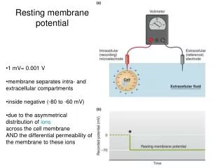
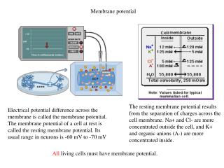
The membrane potential (Vm) is defined as Vm = Vin – Vout Vout = 0 Vin extracellular intracellular Resting membrane potential -60 to –70 mV for neurons Depolarization become more positive Hyperpolarization become more negative
What is membrane potential? Why is it important? How is membrane potential generated? How do we calculate membrane potential? How does membrane potential encode signals? What are the carriers for membrane potential?
The membrane potential results from a separation of positive and negative charges across the cell membrane
I R P V R . Q . Q = P/R I = V/R ’s LAW C Electrical and thermodynamic forces determine the passive distribution of ions C1 C2 Diffusion down Chemical Gradient J R J = C/R V Diffusion down Electrical Gradient C2+ C1+ + + + + + + I + + + + + + I = C/R R
Concept of an Equilibrium Potential for an ionic species: The potential at which the movement of ions across the membrane is in electrochemical equilibrium, i.e. the voltage necessary to result in no net movement of the ionic species across the membrane. Na+ Na+ Na+ Na+ Na+ Na+ Na+ Na+ Na+ Na+ Na+ Na+ Na+ Na+ Na+ Na+ Na+ Na+ +55 mV -90 mV In Equilibrium Out of Equilibrium
gK K+ moves out of cell Positive to EK Negative to EK K+ moves into cell gK Normal current injection Increased gK Voltage response K+ moves out of cell gK K+ moves into cell Maintaining the resting membrane potential Extracellular Intracellular - - - - - - - - - + + + + + + + + + Reversal potential (No net movement of K+) K EK K K K K K K Time Concentration gradient voltage gradient K K K K K K K K K EK -102 -102 mV Time
What is membrane potential? Why is it important? How is membrane potential generated? How do we calculate membrane potential? How does membrane potential encode signals? What are the carriers for membrane potential?
RT F [Na]o [Na]i RT F [K]o [K]i ENa = ln Ek = ln RT 2F [Ca]o [Ca]i ECa = ln RT -F [Cl]o [Cl]i ECl = ln -60 to -75 mV NSCC extracellular ECl = +125 Ca2+ (1.2) ENa = +56 Na+ (150) EK = -102 K+ (3) ECl = -76 Cl- (120) intracellular Na+ (18) K+ (135) Ca2+ (0.1 µM) Cl- (7) Na+,K+-ATPase Resting membrane potentials • Nernst equations for biological ions: Anions: Cl- and proteins Cations: K+ diffusion potential Na+ diffusion potential Ca2+ diffusion potential Na+/K+-ATPase
Important: Ionic concentration differences across cell membranes determines the membrane potential The concentration differences of ions are due to the biophysics of the channels and pumps Guyton, Textbook of Physiology
Nernst Equation E (ion) = RT/zF ln ([ion]outside/[ion]inside) Na+ Na+ Na+ @ 370 C RT/F= (27/z) Convert to log 2.3 x 27/z = 63 Na+ Na+ Na+ Na+ Na+ Na+ @ 0o C = 54 @ 24oC = 59 @ 37oC = 63 E (ion) = 63 log ([ion]outside/[ion]inside)
10 = +73 mv 142 ENa+ +72 0 Vm Membrane Voltage or Potential (mV) - 4 = 89 mv 103 -1 -89 Ecl- -90 Vr (i.e. resting Vm) EK+ -97 = -97 mv 4 140 Time The “Voltage Diagram” ENa+ = 63 log [ ]o/[ ]i ECl- = 63 log [ ]o/[ ]i EK+ = 63 log [ ]o/[ ]i
ENa+ (Equilibrium Potential for Na+) +72 0 Vm Membrane Voltage or Potential (mV) -89 ECl- (Equilibrium Potential for Cl- ) -90 Vr (i.e. resting Vm) EK+ (Equilibrium Potential for K+) -97 Time The “Voltage Diagram” + - - + - + - +
RT F pK[K+]o + pNa[Na+]o + pCl[Cl-]i pK[K+]i + pNa[Na+]i + pCl[Cl-]o Vm = ln -60 to -75 mV NSCC extracellular ECl = +125 Ca2+ (1.2) ENa = +56 Na+ (150) EK = -102 K+ (3) ECl = -76 Cl- (120) intracellular Na+ (18) K+ (135) Ca2+ (0.1 µM) Cl- (7) Na+,K+-ATPase Maintaining the resting membrane potential The Goldman-Hodgkin-Katz Equation: The steady state membrane potential for a given set of ionic concentrations inside and outside the cell and the relative permeability of the membrane to each ion
Three Primary reasons for a net negative potential across the membrane Anion- Anion- Anion- Anion- Anion- Anion- (1) (3) (2) Relative Permeabilities of dominant cations Electrongenic Na/K ATPase High IC [Anions] 3 Na+ EXTRACELLULAR SPACE Na+ + + + + + + + + + + + + + + + + + + + + + + + + + + + + + + + + + + + + + + + + + + + ATPase Lipid Bilayer - - - - - - - - - - - - - - - - - - - - - - - - - - - - - - - - - - - - - - - - - - - - - - - - - - - - - - - - - - - - - - K+ 2 K+ INTRACELLULAR SPACE
Changes in membrane potential due to ion movement • Depolarization: • Initiators: Na+ channels • nonselective cation channels (NSCC) • Na+,K+-ATPase • Terminators: K+ channels • Cl- channels -60 to -75 mV NSCC extracellular ECl = +125 Ca2+ (1.2) ENa = +56 Na+ (150) EK = -102 K+ (3) ECl = -76 Cl- (120) intracellular Na+ (18) K+ (135) Ca2+ (0.1 µM) Cl- (7) Na+,K+-ATPase
What is membrane potential? Why is it important? How is membrane potential formed? How do we calculate membrane potential? How does membrane potential encode signals? What are the carriers for membrane potential?
Action potentials: Signals that travel for long distances through the neuron without losing strength. Types of electrical signals • Graded potentials: Variable-strength signals that lose strength as they travel through the cell. a. Can be depolarizations (Na+ channel) or hyperpolarizations (K+ or Cl- channel) b. Begins on the cell membrane at the point where ions enter from the extracellular fluid (local current or electrotonic current) c. The strength or amplitude is directly proportional to and is determined by the number of charges that enter the cell, which in turn is determined by the number of receptors which are opened. (concentration of the neurotransmitters and density of the receptors) d. The size of the graded potential decreases as it spreads out from its point of origin e. Graded potentials travel through the neurons until they reach the trigger zone, the point where an action potential is generated. Depending on the strength of the graded potential, it either triggers an action potential or dies out (threshold potential). f. Can be summed: spatial summation and temporal summation a. Rapid electrical signals that pass along the axon to the axon terminal. b. Identical to each other and do not diminish in strength when traveling through the cell c. The strength of the graded potential that initiates an action potential has no influence on the action potential as long as it is above threshold. d. All-or-none
Feature Graded Potential Action Potential Type of signal Input signal Conduction signal Where it occurs Usually dendrites and cell body. Axon hillock, initial segment and entire length of axon Types of gated ion channels Mechanically or chemically gated channels Voltage-gate channels Ions involved Usually Na+, K+, and Cl- Na+ and K+ Type of signal Depolarizing (Na+ ) or hyperpolarizing (K+, Cl- ) Depolarizing Strength of signal Depends on initial stimulus; can be summed Is always the same as long as graded potential is above threshold; cannot be summed What initiates the signal Entry of ions through chemically or mechanically gated ion channels Above-threshold graded potential arrives at the integration zone Unique characteristics No minimum level required to initiate a graded potential Two signals coming close together in time will sum Threshold stimulus required to initiate action potential Refractory period: two signals too close together in time cannot sum Initial stimulus strength is indicated by frequency of a series of action potentials Comparison of graded potential and action potential
What is membrane potential? Why is it important? How is membrane potential formed? How do we calculate membrane potential? How does membrane potential encode signals? What are the carriers for membrane potentials?
-10 -10 -10 Voltage (mV) Voltage (mV) Voltage (mV) -100 -100 -100 Na+ K+ Na+/K+ INa,t IK Ih IA INa,p IM Ca2+ ICa,L IC ICa,N ICa,T Voltage-gated ion channels: currents Inward currents Outward currents
Voltage-gated ion channels: structure • Perez-Reyes, Cell Mol Life Sci 56, 660-669, 1999
The structure of mammalian Kv1.2/Kvb2 Long et al, 2005 Science
Voltage-gated ion channels: the superfamily Yu et al., Pharmacol Rev 57: 387-395, 2005
a2 a1s d g b b Voltage-gated ion channels: structure of Ca2+ channels Skeletal muscle L-type Cardiac muscle L-type Bers and Perez-Reyes, Cardiovasc. Res. 42, 339-360, 1999
. . . . . ions 5 mV 20 ms . . ligand . . . Ligand-gated ion channels (ionotropic receptors) Bind to neurotransmitters Receptor channels Mediate fast synaptic transmission Presynaptic terminal Postsynaptic terminal ICa a1B NMDAR AMPAR Ca2+ Ca2+ DAG PLCb4 IP3 Gq/11 Ca2+ mGluR1
GluR1 10% GluR2 GluR3 GluR4 GluR5 GluR6 GluR7 KA1 KA2 NR1 NR2A NR2B NR2C NR2D NR1 NR2 NR2 NH2 NR1 TM1 TM4 TM3 TM2 COOH ionotropic glutamate receptors AMPA: a-amino-3-hydroxy-5-methyl-4-isoxazoleproprionic acid kainate NMDA: N-methyl-D-aspartate Coincidence detector because of voltage dependent Mg2+ block NH2 TM4 NR1 TM1 TM3 TM1 TM3 extracellular TM4 NR2 NR2 Mg2+ TM4 TM3 TM1 TM4 TM1 TM2 NR1 TM3 TM3 TM1 intracellular TM4 COOH
g a b a d NH2 TM1 TM2 TM4 COOH TM3 Cys-loop superfamily Muscle-type Neuronal type Cation channels Nicotinic acetylcholine receptor I, epitheliala9; II, neuronal a7,8; III, neuronal a2–6and b2–4 III-1: a2,3,4,6; III-2, b2,4 III-3, a5, b3; IV, muscle a1, b1,g, d, and e IV-1, a1 IV-2, g, d, e IV-3, b1. 5-HT3 serotonin receptor 5-HT3A, 5-HT3B Anion channels GABAA receptor Glycine receptor subfamilies homo-oligomeric a7 NH2 COOH extracellular TM1 TM2 TM3 TM4 intracellular hetero-oligomeric a4b2 Corringer et al., Annu. Rev. Pharmacol. Toxicol. 40:431-458, 2000 TM4 TM4 TM3 a TM1 g TM1 TM2 TM3 TM2 TM3 TM1 b TM4 TM2 a TM2 TM4 TM2 TM1 d TM3 TM3 TM1 TM4
P2X receptors P2X2 a b P2X3 Cysteine rich extracellular loop P2X5 P2X6 P2X1 P2X4 Plasma membrane P2X7 NH2 Each channel may contain three to six subunits • Activated by ATP • Cation nonselective • ~6.5% of current is carried by Ca2+ C tail length varies Khakh et al., Pharmacol. Rev. 53, 107-118. 2001
GABAA receptor CLC ClC-5 Anion channels
Text books: Chapter 6 Fundamental Neuroscience Chapter 7 Principles of Neural Science