Download
1 / 43
500 likes | 1.61k Vues
Carpal Tunnel Syndrome. Wren V. McCallister, MD Surgery of the Hand & Upper Extremity. Paget 1854. Lectures on Surgical Pathology.

E N D
Carpal Tunnel Syndrome Wren V. McCallister, MD Surgery of the Hand & Upper Extremity
Paget 1854 Lectures on Surgical Pathology “…the median nerve, where it passes under the annular ligament, is enlarged with adhesions to all the adjacent tissues, and induration of both it and them (sic)” “He had ulcerations of the thumb, fore, and middle fingers, which resisted various treatment” Paget J. Lectures on Surgical Pathology. Philadelphia: Lindsay & Blakiston, 1854.
Paget (continued) “…and was cured only by so binding the wrist that the parts on the palmar aspect being relaxed, the pressure on the nerve became and remained well, but as soon as the man was allowed to use his hand, the pressure on the nerve was renewed, and the ulcerations of the parts supplied by them returned”
Putnam (1880) • 37 patients with nocturnal or early am numbness • First description of cardinal symptom of CTS Treatments? Outcome galvanism strychnine cannabis indica …felt “electrified” stopped ALL symptoms just hungry all the time
Marie and Foix (1913) • “hourglass” configuration of nerve nodular thickening, then constriction at the annular ligament • Recommended: if diagnosed early, surgical ”…transection of the ligament could stop the development of these phenomena”
Learmonth (1933) “The median nerve was exposed at the wrist joint. It was compressed between the anterior annular ligament and the arthritic outgrowths of the carpal bones. Scissors were passed under the skin so that one blade was superficial and the other deep to the annular ligament, which was then divided completely.”
Epidemiology of CTS • Incidence of 99 to 148 per 100,0001 • Prevalence from 1% to 10%2 • occupational prevalence: 17% to 61%3 • butchers, grinders, grocery-store workers, frozen-food factory workers (forceful repetitive hand motions, vibration) 1 Palmer DH, Hanrahan LP. Social and economic costs of carpal tunnel surgery. In Jackson DW (ed): Instructional Course Lectures. American Academy of Orthopaedic Surgeons, St, Louis, Mosby 1995, 167-72. 2 Spinner RJ et al. The many faces of carpal tunnel syndrome. Mayo Clin Proc 64:829-36, 1989. 3 Hagberg M et al. Impact of occupations and job tasks on the prevalence of carpal tunnel syndrome. Scand J Work Environ Health 18:337-45, 1992.
4th-5th decade (82% > 40yo) • Female:Male 3:1 • ~50% have bilateral CTS • up to 38% contralateral wrists: Asx with abnormal NCV • ~400,000-500,000 CTR per annum (USA)1 • economic cost ~ $2 billion • worker’s comp cost 3X other workers • worker’s comp cost 5X non-workers 1 Palmer DH, Hanrahan LP. Social and economic costs of carpal tunnel surgery. In Jackson DW (ed): Instructional Course Lectures. American Academy of Orthopaedic Surgeons, St, Louis, Mosby 1995, 167-72.
What about Work? • 22 epidemiologic studies to identify risk factors • OR from 1.7 to 34 • consistent evidence to support association • repetitive motion and forceful motion • non-neutral wrist postures, vibration • cold temperatures • did not control for force/repetitive motion • synergy for > 2 risk factors • dose-response (suggested but not proven) • No established cause and effect Hales TR, Bernard BP. Epidemiology of work-related musculoskeletal disorders. Ortho Clin N Amer 27(4):679-709, 1996.
Stevens, Neurology 2001 No causal relationship Rates ~ general population
Other risk factors • Obesity • Hypothyroidism • Diabetes (prevalence 14%-30% with neuropathy) • Pregnancy (~50% prevalence) • Renal disease • Inflammatory arthritis • Acromegaly • Mucopolysaccharidosis • Genetics (twin study) • Age (>50) • Smoking
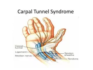
Anatomy of the Carpal Tunnel FCR FPL FDS ----- FDP
Carpal Tunnel Topography • Proximal border = palmar wrist crease • Distal border = Kaplan + ring finger axis Thenar motor branch Kaplan’s cardinal line: distal TCL thenar branch superficial arch Superficial palmar arch
Median Nerve • Originates lateral and medial cords of brachial plexus • Contributions from C6, C7, C8 & T1 (± C5) • Motor fascicles (radially oriented) • Thenar branch variations
Disturbed axoplasmic flow Endoneural edema Impaired neural circulation Diminished nerve elasticity Decreased gliding Pathophysiology
Chronic CTS • Classification • Early • mild sx (night, activity) • < 1 year duration • no gross morphologic changes in nerve • Intermediate • numbness, paresthesias (min. thenar atrophy) • chronic changes in median nerve (edema) • reversible with decompression
Chronic CTS • Advanced • marked sensory changes • thenar motor weakness • chronic pathologic changes in median nerve • endonerual edema, intraneural fibrosis, partial demyelination, axonal degeneration • some changes irreversible
Pathophysiology • Clinical stages: • magnitude and duration of compression • Normal subjects • carpal tunnel pressure = 2.5mmHg (neutral) • CTS subjects • carpal tunnel pressure = 32mm Hg (neutral) • 94-110mmHg with wrist flexion/extension • epineural edema (<2 h), endoneural edema
Pathophysiology • Symptom relief after decompression • Immediate • restore intraneural blood flow in normal nerve • Days-weeks • decreased intraneural edema • Months • remyelination and axonal regeneration
History • Common presentation • intermittent pain and paresthesias in the median nerve distribution • nocturnal paresthesias (cardinal Sx) • with time, thenar atrophy • weak grip, fatigue with repetitive activity • sensory-sparing CTS • can be clumsiness/weakness of hands • “shake test”
Differential Diagnosis • C6, C7 radiculopathy • Thoracic outlet syndrome • Proximal median nerve entrapment • Traumatic injury at the level of the wrist • handcuff neuropathy • Double crush syndrome • Upton, McComas (Lancet 1973) • 81/115 patients with median/ulnar nerve sx also had cervical nerve root lesion
Physical Exam • Clinical findings • wasting of thenar eminence • weakness of APB (most sensitive motor sign) • palmar abduction / thumb supination • weakness of opponens pollicis • Skin examination • ulcerative, necrotic or bullous lesions • digital anhydrosis, alopecia, nail change (rare)
Physical Exam - Sensory • Threshold testing • Semmes-Weinstein monofilament or vibrometry • Preferred method of testing sensibility • Vibrometry more sensitive, less practical • Innervation density testing • Static two-point discrimination • “slow” adapting fibers • Moving two-point discrimination • “fast” adapting fibers
Two-point discrimination Moberg 1958 Static (nl < 6mm) and Moving (nl = < 3mm) Abnormal = severe nerve compression
Semmes-Weinstein • Von Frey hairs (1898) • Five selected thresholds: • normal (2.83), light touch (3.61), protective (4.31), loss of protective (4.56), loss of deep pressure (6.56) • Abnormal > 2.83 (eyes closed)
Vibrometry • Dellon 1980 • Biothesiometer (shown) • Evaluates “fast” adapting fibers • More expensive, cumbersome than monofilament testing
Ten Test • “10 test” (Strauch, Plast Rec Surg 1997) • Patient ranks moving LT from 0-10 compared to normal contralateral area • Useful adjunct for serial examinations • Correlates with SW monofilament testing
Physical exam • Provocative testing • ALWAYS, test sensibility first ! • many described, all based on same concept • stress a compromised median nerve to recreate Sx • 3 most commonly used tests • Phalen’s test, Tinel’s test, compression test • Tourniquet test • high false (+) rate
Phalen’s test • Described in 1951 • Originally: rested elbows on table • better without elbow flexion • Median nerve trapped b/n proximal TCL and underlying flexor tendons & radius • “reverse” Phalen’s maneuver • Abnormal = reproduce Sx in 30-60 sec • Limitations • decreased wrist motion, severe CTS • wide variation in reported sensitivity (10%-80%) and specificity (40%-100%)
Tinel’s Sign • Gently tapping along the median nerve at the wrist • Abnormal = tingling in median nerve dist. • Careful to tap “gently” • Phalen reported 60%-73% of patients with CTS had a Tinel’s sign present • Wide range of sensitivity (26%-79%) and specificity (40%-100%)
Durkan Compression Test • Gentle pressure directly over carpal tunnel paresthesias in 30 seconds or less • Better for wrists with limited motion • Highest sensitivity/specificity of all physical exam tests
Summary of Tests Test Sensitivity Specificity Phalen’s 75% 62% Tinel’s 64% 71% Compression 87% 90% S-W monofilament 65% 42% Vibrometry 87% ?
Electrodiagnostic Tests • NOT the gold standard • Benchmark for validity testing in CTS • how physical exam tests are evaluated for accuracy • Diagnostic bias • selection criteria for application of test • different methods of performing tests • patient selection differs from study to study • Spectrum bias • use of asymptomatic controls for sens/spec • goal of test = identify those with disease in a pool of patients with symptoms c/w the disease
Electrodiagnostic Tests • Latency and conduction velocity • reflect only the healthiest myelinated axons • large fibers only (not pain / temperature) • can be normal in early stages of compression • dynamic ischemia • EMG • can distinguish functional symptoms • normal study except for submaximal voluntary MUP recruitment
Electrodiagnostic tests • Abnormal = across the wrist: • distal motor latency > 4.5ms • sensory latency > 3.5ms • However: • 8-22% of patients with (-) electrodiagnostics and (+) clinical signs improve with CTR • electrodiagnostics (+) for Asx, (-) for Sx
Diagnosis of CTS • Consensus Statement (Am J Pub Health 1998) • (-) ED test, (+) classic sx = ? If CTS • (+) ED test, (-) symptoms CTS • Szabo 1999 • night pain, (+) SW, (+) Durkan’s, (+) Hand diagram = 86% probability of CTS • all test above (-) = 0.68% probability of CTS • ED tests did not add to diagnostic power • CTS is a clinical diagnosis • ED tests can help: • identify peripheral neuropathy • locate other sites of compression • establish severity
Non-operative Treatment • Mild to moderate disease • key is denervation of ABP • Splinting (nocturnal, neutral) • Oral agents • NSAIDs, Vitamin B6 (?) • Neither effective in isolation • Steroid injection • 80% relief short-term, ~10-20% @ 1.5 years • (+) response predictive of success with surgery • dexamethasone safest
Non-operative Treatment • JBJS Evidence-Based Orthopaedics* • “Decompressive Surgery Was Better Than Steroid Injection for Symptomatic and Neurophysiologic Outcomes in Carpal Tunnel Syndrome” • PRCT, ED-proven CTS, 20wk f/u • All injection patients had improvement • Pain, NCV better with surgery (not grip) *McCallister, Trumble JBJS (Am) 2006
Non-operative Treatment • Therapy • iontophoresis + splint ? > NSAIDs + splint • ultrasound is equivocal • Activity/ergonomic modification • Exercises • aerobic exercise ? • yoga ? short-term benefit • tendon and nerve gliding* • 43% failure versus 71% if not done @ 2y f/u *Rozmaryn et al, J Hand Ther 1998
Non-operative Treatment • No benefit: • magnets • laser • acupuncture • chiropractic
Operative Treatment • Indicated when non-operative treatment has failed or thenar motor denervation • Minimally-invasive Endoscopic Carpal Tunnel release • Evidence supports success of Endoscopic Carpal Tunnel release and suggests earlier return of function compared to open release
Summary • CTS is a clinical diagnosis • ED are confirmatory, if not required (L&I) • No cause and effect vis-à-vis work • Non-operative treatment early • Operative treatment • if denervation of APB • failure of non-operative treatment