Download
1 / 33
410 likes | 1.24k Vues
Chapter 3: Understanding the Brain and Brain Injury. Module Objectives. Identify basic brain structures and functions. Describe brain-behavior relationships. Describe how an injury to the brain can result in various behaviors and challenges. Introduction.

E N D
Module Objectives • Identify basic brain structures and functions. • Describe brain-behavior relationships. • Describe how an injury to the brain can result in various behaviors and challenges.
Introduction The brain is the main organ of learning. • It makes it possible for us to think, communicate, act, behave, move about, and create.
Mechanisms of Traumatic Brain Injury After a sudden jolt or bang, the result can be… • Coup-Contracoup: Injury at the site of impact and on the opposite side from the movement of the brain against the skull (either front to back or side to side) Courtesy Centre for Neuro Skills
Mechanisms of Traumatic Brain Injury After a sudden jolt or bang, the result can be… • Diffuse Axonal injuries: Delicate nerve tissues rip, tear, and stretch • Swelling: Brain tissue swells preventing blood and CSF circulation • Hematoma: Accumulation of blood causing pressure • Hydrocephalus: Blockage of CSF causing pressure • Anoxia & Hypoxia: Oxygen deprivation from suffocation, drowning, blood loss, or cardiac failure that kills brain cells Courtesy Centre for Neuro Skills • Hemorrhages: Major bleeding from when the brain rubs against the inside of the skull, which is ragged with sharp bony ridges
When the Brain is Injured A brain injury is often the result of two injuries: • A “primary injury” caused by the initial blow or insult to the brain • A “secondary injury” caused by the swelling, bleeding, compression and contusions (bruises) to the brain.
Severity of Brain Injuries Glasgow Coma Score (GSC) • Is a measure of brain injury severity. • Measures Eye Response + Verbal Response + Motor Response = Total Score • Scores range between 3 and 15 • The lower the score, the more severe the brain injury
Severity of Brain Injuries Post concussion symptoms of cognitive and psychiatric nature that may or may not persist include: headache changes in personality dizziness memory problems vomiting depression sleep disturbance difficulty problem solving irritability diminished attention span
Anatomy of the Brain The brain . . . • Is a soft organ, like the consistency of gelatin • Weighs less than 1 lb. at birth and grows to about 3 lbs. • Sits inside a rough and bony skull and is bathed in a cerebrospinal fluid (CSF) • Receives oxygen and glucose through a sophisticated system of blood vessels that carry blood to and from the heart
Anatomy of the Brain Three membranes or meningescover the brain: • The outer dura materor hard matter, which is like a heavy plastic covering. • The arachnoid, which is like a spider web that bridges the brain's many wrinkles and folds. • The pia mater or tender matter, which molds around every tiny crook and crevice on the brain's surface. • Between the pia mater and the arachnoid, there is 145cc of cerebrospinal fluid. Scalp Skull Dura Mater Arachnoid Pia Mater
Anatomy of the Brain There are four ventricles which make, store, and circulate cerebrospinal fluid. • The fluid helps cushion the brain and protect brain tissue when swelling occurs.
Neurons • Neurons:the billions and billions of tiny brain cells making up the nervous system • Glial ("glue"): non-communicating cells support and nourish the neurons. • Three main parts of the neuron:
Neurons • The neurons communicate with each other via a unique“electro-chemical”process. • Neurotransmitters are chemical messengers that relay the electrical signal of one nerve cell to the next. • Neurochemical transmitters leap the synaptic gaps. • After a person sustains a brain injury, many of the neuron pathways may be torn apart or stretched so that information processing is no longer possible.
Brain Stem • Midbrain • Alertness & arousal • Elementary forms of seeing & hearing • Pons • Facial movement & sensation, hearing, & coordinating eye movements • Medulla • Basic living functions • Vital to life and death • Controls involuntary functions like breathing, heart-rate, blood pressure, swallowing, vomiting and sneezing.
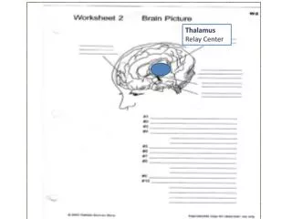
diencephalon • Thalamus • Major relay station for incoming and outgoing sensory information • The input for every sense (except smell) travels through the thalamus • Hypothalamus • Control center for hunger, thirst, sexual response, endocrine level & temperature regulation. • Controls complex responses like anger, fatigue, memory and calmness.
Limbic System • Limbic System • Houses basic elemental drives, emotions and survival instincts. • Injury to the limbic system can result in serious problems with basic emotional perceptions, feelings & responses. • Behavior and mood can be very erratic
Limbic System Amygdala Fight or flight structure The front door to our emotions When perceptions reach the cerebral cortex, it is transmitted to the amygdala to be evaluated for emotional content Hippocampus Associated with memory functions Injury can result in problems with short term memory, and turning short term memories into long term memories Disrupts the encoding and retrieval of long term memory
The Cerebral Cortex • Cerebral Cortex:the most complicated structural component of the brain • Made up of two hemispheres: the right hemisphere and left hemisphere • Dedicated to the highest levels of thinking, moving, and acting. • Each hemisphere is divided into four lobes – frontal, parietal, temporal, and occipital • The cortex is full of wrinkles and folds. • If you took out and flattened the cortex, it would be the size of a pillowcase. Right Hemisphere Left Hemisphere
The Cerebral Cortex • The two hemispheres of the brain have unique ways of processing information. • The right hemisphere is more holistic, visual–spatial, and intuitive. • The left hemisphere processes language and is more linear, verbal–analytic, and logical. • The cerebral hemispheres control opposites sides of the body. • The cerebral hemispheres communicate to each other a thousand times a second through the corpus collosum (the 4 inch long, pencil thick band of complex nerve fibers).
Lateralized Skills of the Brain • The brain is divided into two hemispheres • The left hemisphere controls the right side of the body. • The right hemisphere controls the left side of the body • The two hemispheres control input and regulate output
Lobes of the Brain Parietal lobe Frontal lobe Occipital lobe Temporal lobe Cerebellum
Lobes of the Brain continued • The lobes are interconnected by complex neural fibers, which relay impulses and information to and from the cortex. • Each lobe has a right and left side.
Brain & Behavior Relationships Parietal Lobe Frontal Lobe Initiation Problem solving Judgment Inhibition of behavior Planning/anticipation Self-monitoring Motor planning Personality/emotions Awareness of abilities/limits Organization Attention/concentration Mental flexibility Speaking Sense of touch Differentiation of size, color, shape Spatial perception Visual perception Occipital Lobe Visual perception and input Reading (perception and recognition of printed words) Cerebellum Coordination Balance Skilled motor activity Temporal Lobe Brain Stem Memory Hearing Expressive and receptive language Comprehension of language Musical awareness Organization &sequencing skills Breathing Heart rate Arousal/Consciousness Sleep/wake functions Attention/concentration
Vulnerable to injury since they sit just inside the front of the skull near a rough bony area Have extensive connections with many brain regions, especially with the parietal lobe and the limbic system (emotions). Includes the motor strip Sends signals to the muscles of the body, telling them what to do Frontal Lobes Prefrontal cortex:located at the very front part of the frontal lobes Helps hold information in memory for several minutes (referred to as working memory) Regulates emotional responses, motivation, executive functions, working memory Responsible for teaching a person to learn from consequences
Frontal Lobes Motor Strip Prefrontal Cortex
Frontal Lobe Injury Injury damages an individual's ability to . . . • Synthesize signals from the environment • Assign priorities • Make decisions • Initiate actions • Attend to tasks • Control emotions • Behave and interact socially • Make plans
Frontal Lobe Injury in Children • Prefrontal lobe injuries in young children sometimes go unnoticed • Parents and teachers typically function as their frontal lobes—they organize, plan, and direct their children’s lives. • As the child gets older and enters early adolescence, they are expected to be more independent and learn to manage themselves over time. • In the child with a brain injury, the capability for more independent frontal lobe functioning has been diminished.
Parietal Lobe • Situated behind the frontal lobes • Includes the primary sensory cortex which is posterior to the motor strip. • The first part of the brain to consciously register physical sensations. • Regulates responses to touch, heat, cold, pain, and body awareness Sensory Strip
Parietal Lobe Injury • When one side of the lobe is injured, a person may not recognize that anything is wrong with movement on the other side of the body. • Even more complex functions like attention can be affected by damage to the parietal lobes.
Occipital Lobe • Located in the lower back part of the brain • The primary visual center of the brain • Involves the visual cortex • Connected to the eyes by optic nerves • Optic nerves carrying signals meet at a "crossing" called the optic chiasm • The left optic track carries signals from the right–side field of vision, and the right optic track takes signals from the left so that both sides of the brain "see" the same thing. • Most of what a person "sees" derives its meaning from prior learning and symbolic representations.
Temporal Lobes • Rest on both sides of the brain • The centers for language & hearing • Broca’s Area • located in the lower portion of the motor cortex in the left frontal–temporal lobe • Controls muscles of the face and mouth and enables the production of speech • Wernicke’s Area • located left temporal–parietal lobe • Governs a person’s understanding of speech • With their connections to the hippocampus, the temporal lobes help in the long–term storage of permanent memories.
Temporal Lobes Broca’s Area Wernicke’s Area