Download

1 / 46
780 likes | 2.2k Vues
Management of head injury. epidemiology. Head injury is a leading cause of morbidity and mortality and it is the most common cause of death among all patient suffering of traumatic injury. Motor vehicle accident are the most frequent cause of traumatic brain injury peaking in 15-24

E N D
epidemiology Head injury is a leading cause of morbidity and mortality and it is the most common cause of death among all patient suffering of traumatic injury. Motor vehicle accident are the most frequent cause of traumatic brain injury peaking in 15-24 Childe abuse is a common cause of head injury in very young infants Falls is the most common source of head injury in patient above 80
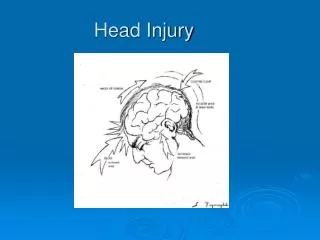
Pathophysiology • Head injuries include both injuries to the brain and those to other parts of the head, such as the scalp and the skull. • Brain injury is divided into primary and secondary • Primary: occurs at the time of brain injury( direct result of trauma ) or immediate there after eg:(skull fracture ,laceration of the brain , hemorrhage in or around the brain) • Secondary ( after the primary ): denotes the brain response to injury eg:(cellular ischemic injury and cerebral edema )
Primary brain injury • Primary brain injury could be caused by penetrating or blunt trauma . • The brain is much more sensitive to diffuse angular application of force than focal linear one.
Secondary brain injury • They occur from subcellular to macroscopic level • Tissue ischemia due to the fact that Cerebral blood flow drop concededly in the early face after traumatic brain injury. • Cerebral edema occur due to cytotoxic ( BBB is intact) and vasogenic ( BBB is broken ) mechanism and acts as an additional mass which increase intracranial pressure .
Clinical assessment • The basic management is divided into: • initial resuscitation(a,b,c,d,e) • primary neurologic survey(glasgow coma scale+ pupils+gross motor) • search for lesions that require immediate surgical management • identification and management of cerebral edema and increased intra cranial pressure.
All patient with sever brain injury may require intubation purely on the basis of a reduced ability to protect the airway and maintain ventilation. • Restoring and maintaining adequate blood pressure is important • data base research demonstrate that a single episode of hypotension following brain injury doubles the risk of dying compared to patients who never had an episode.
The score range from 3-15. • A score between 3-8 indicate severe brain injury . • A score between 9-12 indicate moderate brain injury. • A score between 13-15 indicate mild closed head injury.
In addition to determining the GCS the primary neurological survey include an assessment of the pupil and gross motor function . • The mass of hematoma may produce uncle herniation with unilateral pupillary dilatation and contralateral hemiparesis and these indicate a life threatening intra cranial hemorrhage is present.
Sign and symptoms • Presentation varies according to the injury. A patient may present with or without neurologic deficit. • Common symptoms of head injury include: coma, confusion, drowsiness, personality change, seizures, nausea and vomiting, headache and a lucid interval, during which a patient appears conscious only to deteriorate later. • When we have a skull fracture : leaking cerebrospinal fluid and is strongly indicative of basilar skull fracture. an eye that cannot move or is deviated to one side. wounds or bruises on the scalp or face. Basilar skull fractures, those that occur at the base of the skull, are associated with Battle's sign, a subcutaneous bleed over the mastoid hemotympanum, and cerebrospinal fluid rhinorrhea and otorrhea.
Diagnostic imaging • All patient with moderate and sever brain injury should have a CT scan of the head . Also with mild with risk factor such as the use of anticoagulants. • Plain skull X ray is not advised as substitute for CT. • MRI maybe useful in assessing cervical and intracranial vasculature and helpful in accurate prognostication of injury outcome
Conventional angiography ,CT scan ,and MRA are potential consideration in case of cervical intra cranial vascular injury • Conventional angiography is the gold standard and offers endovascular treatment. • After the head CT scan the clinical and radiographic information should be synthesized to formulate a plan either to operate or evaluate for intra cranial hypertension or to observe clinically for worsening of neurological status
Clinical injury pattern and herniation syndrome • Immediate recognition and treatment of these syndromes is essential for patient survival. • Subfalcine herniation occur when part of the cerebrum is forced from one side to an other under the falxcerebri. • There is a general correlation between the degree of Medline shift , the depression in the level of consciousness and the severity of the injury.
Uncal herniation • Occur when the uncus of the temporal lobe is shifted medially • It can compress the oculomotor resulting in loss of it’s parasympathetic function • With more sever or prolonged compression the extra oculor muscle function is also lost leading to an eye that is deviated anteriorly and laterally. down and out position in the affected eye • if the uncus is forced further medially it compress the cerebral peduncle producing counter lateral hemiparesis
Tonsillar herniation • Occur when the intra cranial content particular the content of the posterior fossa are forced out of the foramen magnum • The tonsils of the cerebellum are pushed downward and compress the medulla oblongata leading to respiratory depression and death
The cushing triad • This is physilogical response of high ICP • Bradycardia • Hypertension • Respiratory irregularity • Intubation and mechanical ventilation often obscure the respiratory part of the triad
Specific injuries & surgical management • Skull fracture • They are usually categorized into open or closed( non- penetrating) depressed or non depressed and basilar or convexity varieties • Depressed fracture can tear the Dura or lacerate the cortex of the brain and they are usually considered for operative repair • Skull fractures that are not depressed usually do not require repair • Basilar skull fractures can damage the the vasculature and crainial nerves and can lead to a CSF leak and meningitis • Clinically one may suspect a basilar skull fracture in the presence of Battle’s sign or Raccoon eyes
Battle’s sign Raccoon eyes
Epidural hematoma • The bleeding occures on the outside of the dura and collects in and expands the potential epidural space between the dura and the bone • The classic example is laceration of the middle meningeal artery by the temporal bone because of fall or blow on the temple • The hematoma can compress the adjacent brain to the point of serious injury and death • venous epidural hematoma are less likely to produce life threatening compression • Distinguishing between the two types is location (temporal vs non temporal) ,size (small vs large) ,and the rate of exchange (slow vs fast)
The classic presentation of a tamporal epidural hematoma is a patient suffering from a blow of the temple and a brief loss of consciousness . This is fallowed by a lucid interval during which the patient appears to be neurologically well • during this time either the epidural hematoma expands slowly enough for the compensatory mechanism of the brain to maintain consciousness or the hemorrhage stops temporarily • Subsequently either due to rebleeding or exhaustion of the compensatory mechanisms a rapid decline in the level of consciousness and associated with other element of uncal herniation
Epidural hematoma has a lens shaped appearance and limited by the suture lines • CT image is also helpful in assessing the impact of the hematoma on the brain such as the degree of the midline shift or compression of the nearby structures • Management of epidual hematoma is determined by lesion size ,location and time from injury to diagnosis • Generally, an epidural hematoma more than 1cm in depth is considered for surgical management( crainiotomy) • When the diagnosis has been delayed, it is considered safe to observe patients with epidural hematoma of modest size (1cm) since the bulk of the hematoma expansion occures within the first 24-36 houres
In contrast , a 1cm thick temporal epidural hematoma in a patient only 30 minutes out from injury should be strongly considered for emergent surgical removal of the hematoma
Acute subdural hematoma • The blood collects between the dura and the arachnoid membrane , in the potential subdural space • Shaking (acceleration-deceleration) injury leading to tearing of bridging veins in the subdural space. In young children, this may result from child abuse. • Most patients present with a significantly depressed level of consciousness and may have other finding related to the compressive mass of the hematoma ( headache confusion sezures )
It has more of a crescent shape and there is no barrier to it’s spreading over the hemispheric surface of the brain • Associated intracerebral hemorrhage and edema is often evident beneath the acute subdural hematoma • Acute subdural bleeds have a high mortality rate, higher even than epidural hematomas and , because the force (acceleration/deceleration) required to cause them causes other severe injuries as well. The mortality rate associated with acute subdural hematoma is around 60 to 80%. • Management of the acute subdural hematoma involves emergent operative removal by craniotomy for lesions more than 1cm thick • Acute brain swelling during the surgical procedure must be expected • Bleeding source usually difficult to stop and often best managed with gentle packing of hemostatic materials rather than aggressive exploration
Medical management of elevated intracrainialpressure ( osmotic diuritucs and hypervent. Which will dec. PaCo2 which will lower ICP) and the underlying brain injury is an essential part of patient care • Outcomes are often poor with mortality of 50%-90%. Survivors often have significant disabilities • In elderly the subdural hematoma can present several months after injury due to the space caused by brain atrophy and they are labeled as chronic subdural hematoma and outcomes post surgery are much better than acute subdural hematoma
Penetrating brain injury • Usually produce a focal injury pattern specific to the site of penetration • Gunshot wound that pass through the ventricular system are most often fatal • Management of less severe penetrating injury revolves around manging the potential infection ,complication of injury and repairing the breach of the skull
Subarchnoidhemorrage • is bleeding into the subarachnoid space—the area between the arachnoid membrane and the pia mater surrounding the brain • Trauma is most common fallowed by cerebral aneurism • aneurysm is classically associated with the sudden onset of a severe headache, traumatic subarachnoid presentation all depends on the mechanism of injury, the severity of the overall injury and other associated traumatic injuries to the brain. • other types of traumatic hemorrhages, such as epidural hematoma or subdural hematoma, often require surgical removal, bleeding into the subarachnoid space spreads out into the cerebrospinal fluid and thus does not cause a mass that requires evacuation thus does not require any specific treatment
Traumatic intraventricular hemorrhage • bleeding in the ventricular system . Primary IVH is uncommon Intraventricular hemorrhage more commonly occurs in the setting of intracerebral hemorrhage or subarachnoid hemorrhage (secondary IVH). • Management ??
Traumatic IntraparenchymalHemorrhage • bleeding into the tissue of the brain caused by trauma • An intraparenchymal hemorrhage rarely occurs alone so the presenration will vary • smaller hemorrhage often require no specific treatment other than the standard treatment of any trauma victim, very large hematomas may require surgical removal.
Concussion • Is an immediate and transient loss of consciousness or normal mentation after head trauma often associated with period of amnesia • The severity of concussion appears to be proportional to the duration of amnesia particularly the anterograde amnesia
Management for isolated concussion in the ER includes a period of observation for at least 2 hours • Long term outcome are generally good however 25% of patients report an increased incidence of headache and memory difficulties even several months later • post concussive syndrome include depression, anxiety, memory loss , headache , emotional lability , insomnia • Treatment is reassurance
Diffuse axonal injury • Occurs when the axons become sheared off between the boundary between gray and white matter during rapid brain acceleration or deceleration • it does not generally cause specific, focal neurological symptoms. However, it can often contribute to a depressed level of consciousness • contribute to the long-term neurological disability cuz axons cannot regrow • patients who have severe DAI often remain in coma or a persistent vegetative state. • The CT of a patient with diffuse axonal injury may be normal despite the patient's presentation with a profound neurological deficit. With CT, diffuse axonal injury may appear as ill-defined areas of high density or hemorrhage however MRI is better • Management is primarily medical
Medical management after traumatic brain injury • Seizure management • Homeostasis • Management of increased intracranial pressure by (venous drainage , CSF drainage, sedation, osmotic agent and diuretics, hyperventilation ,barbiturate, hypothermia , surgical decompression