Head Injury
Head Injury. Dr Malith Kumarasinghe MBBS (Colombo). Head Injuries:. Account for about one half of all trauma deaths Survivors range from baseline function to severe morbidity Even “minor” head injury can have severe impact As with most trauma, broken down into blunt and penetrating.


Head Injury
E N D
Presentation Transcript
Head Injury Dr Malith Kumarasinghe MBBS (Colombo)
Head Injuries: • Account for about one half of all trauma deaths • Survivors range from baseline function to severe morbidity • Even “minor” head injury can have severe impact • As with most trauma, broken down into blunt and penetrating
Physiology of Nervous System • Cerebral Blood Flow (CBF) = 50 – 55 ml/100g of brain tissue/minute • Main Arterial Pressure (MAP) = Diast. P + 1/3 Pulse P • Intracranial Pressure (ICP) = 10 mm Hg • Cerebral Perfusion Pressure (CPP) = 50 -150 mm Hg • CPP = MAP – ICP
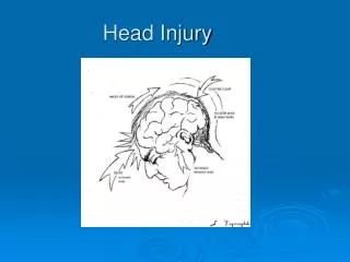
Injuries to the Brain & Skull • Scalp injuries • Skull injuries • Brain injuries
Scalp Injuries • Scalp has many blood vessels so injury may bleed profusely. • Control bleeding with direct pressure. • Don’t apply pressure when there is possible skull injury.
Cephalhaematoma • It is a subperiostealhaematoma most commonly lies over one parietal bone. • It may result from difficult vacuum or forceps extraction Management: - It usually resolves spontaneously. - Vitamin K 1 mg IM is given.
Mechanism of injury in head trauma • Direct trauma by compression or crushing. • Acceleration-Deceleration Injuries • when a person falls backwards onto a hard floor, the back of the person’s head hits the floor and stops. The brain, however, is still moving until it strikes the inside of the skull. If the brain gets bruised, there is bleeding, also called a hemorrhage. This bleeding causes further damage to the brain. • The skull does not need to strike an object in order for the brain to get injured. There are many situations in motor vehicle crashes where the forces are transmitted through the brain without the skull hitting the dashboard, windshield, steering wheel or window. • Coup/Contrer-CoupInjuries:Related to acceleration-deceleration injuries(e.g injury to temporal lobe in contralateral temporal trauma)
Head Trauma 1-Skull fractures. 2-Extradural ,subdural & subarachinoid. 3-Cerebral contusion& intraventricular Hge. 4-Diffuse Axonal Injury (DAI). 5-Related Brain edema & herniation.
Skull Fracture • A skull fracture is a break in the skull bone and generally occurs as a result of direct impact . • Uncomplicated skull fractures themselves rarely produce neurologic deficit, but the associated intracranial injury may have serious neurologic sequelae.
Four major types of skull fractures may occur: • (1) linear, • (2) depressed, • (3) diastatic, • (4) basilar.
Linear fractures, the most common Involve a break in the bone but no displacement, and generally no intervention is required. These fractures are usually the result of low-energy transfer due to blunt trauma over a wide surface area of the skull. The fracture involves the entire thickness of the skull. Generally, these fractures are of little clinical significance unless they involve a vascular channel, a venous sinus groove, or a suture. Complications include epidural hematoma, venous sinus thrombosis, and suture diastasis.
Depressed skull fractures CechPetr
A fracture is clinically significant and requires elevation when a fragment of bone is depressed deeper than the adjacent inner table. Depressed fractures may be closed or compound (open). Compound fractures may be exposed when they are associated with a skin laceration or when the fracture extends into the paranasal sinuses and the middle-ear structures.
Diastatic Skull Fracture Diastatic fractures occur along the suture lines and usually affect newborns and infants in whom suture fusion has not yet happened. In this type of fracture, the normal suture lines are widened
Basilar skull fractures Basilar fractures are the most serious and involve a linear break in the bone at the base of the skull. Most basilar fractures occur at 2 specific anatomic locations—namely, the temporal region and the occipital condylar region. These fractures are often associated with dural tears, of which cerebrospinal fluid (CSF) rhinorrhea and otorrhea are known complications.
Basilar skull fractures • Most basilar fractures occur at 2 specific anatomic locations — namely, the temporal region and the occipital condylar region.
Epidural Hematoma • An epidural hematoma is usually associated with a skull fracture. It often occurs when a direct impact fractures the calvarium . • The fractured bone lacerates a dural artery (middle meningeal artery) or a venous sinus. • On CT, the hematoma forms a hyperdense biconvex mass. It is usually uniformly high density but may contain hypodense foci due to active bleeding. • Comment on midline shift .
Natasha Richardson March 2009
Clinical features • Patients may have external evidence of head injuries such as scalp lacerations, cephalohematoma, or contusions. • no loss of consciousness, brief loss of consciousness, or prolonged loss of consciousness. • The classic lucid interval occurs in 20-50% of patients with EDH. • Initially, the concussive force that caused the head injury results in an alteration of consciousness. • After recovering consciousness, the EDH continues to expand until the mass effect of the hemorrhage itself results in increased intracranial pressure, a decreased level of consciousness, and a possible herniation syndrome.
severe intracranial hypertension, a Cushing response may occur. • The classic Cushing triad involves systemic hypertension, bradycardia, respiratory depression.
Treatment • treatment options for these patients are • (1) immediate surgical intervention • (2) initial, conservative, close clinical observation with possible delayed evacuation.
Subdural Hematoma • Deceleration and acceleration or rotationalforces that tear bridging veins can cause an acute subdural hematoma so it occurs in cases of wide subdural space(old age & children) • Causes of subdural are :in minimal trauma in old age, child abuse and ventricular decompression, may occur in patients receiving anticoagulants or patients with a coagulopathy condition. • The blood collects in the space between the arachnoid matter and the dura matter, Because the subdural space is not limited by the cranial sutures, blood can spread along the entire hemisphere and into the hemispheric fissure, limited only by the dural reflections . • We have 3 major types : Acute, subacute & chronic
Crescent shaped;Hyperdense, may contain hypodense foci due to serum, CSF or active bleeding Acute Subdural Hematoma
In children, subdural hematomas occurring along the posterior interhemispheric fissure and the tentorium have been described as common findings following violent nonaccidentalshaking • (ie, shaken baby syndrome) .
Clinical Features Clinical presentation depends on the location of the lesion and the rate at which it develops. Often, patients are rendered comatose at the time of the injury. A subset of patients remain conscious; others deteriorate in a delayed fashion as the hematoma expands.
Treatment significant acute traumatic subdural hematoma requires surgical treatment, temporizing medical maneuvers can be preoperatively used to decrease intracranial pressure. These measures are germane for any acute mass lesion As with any trauma patient, resuscitation begins with the ABCs
Short-acting sedatives and paralytics should be used only when needed to facilitate adequate ventilation or when elevated intracranial pressure is suspected. The patient should also be mildly hyperventilated Administer anticonvulsants to prevent seizure-induced ischemia and subsequent surges in intracranial pressure. Do not give steroids, as they have been found to be ineffective in patients with head injury.
Subacute Subdural Hematoma • Subacute SDH may be difficult to visualize by CT because as the hemorrhage is reabsorbed it becomes isodense to normal gray matter. • A subacute SDH should be suspected when you identify shift of midline structures without an obvious mass. • Giving contrast may help in difficult cases because the interface between the hematoma and the adjacent brain usually becomes more obvious due to enhancement of the dura and adjacent vascular structures. • Some of the notable characteristics of subacute SDH are:- Compressed lateral ventricle& or midline shift - Effaced sulci .
Subacute subdural hematomas are defined arbitrarily as those that present between 4 and 21 days after injury.
Chronic Subdural Hematoma • Chronic SDH becomes low density as the hemorrhage is further reabsorbed. It is usually uniformly low density but may be loculated. Rebleeding often occurs and causes mixed density and fluid levels.
Chronic subdural hematomas are arbitrarily defined as those hematomas presenting 21 days or more after injury.
Subarachnoid Hemorrhage • A subarachnoid hemorrhage occurs with injury of small arteries or veins on the surface of the brain. The ruptured vessel bleeds into the space between the pia and arachnoid matter. The most common cause of subarachnoid hemorrhage is trauma . • In the absence of significant trauma, the most common cause of subarachnoid hemorrhage is the rupture of a cerebral aneurysm.
Causes • Bleeding from an arteriovenous malformation (AVM) • Bleeding disorder • Bleeding from a cerebral aneurysm • Head injury • Unknown cause (idiopathic) • Use of blood thinners
SAH classification Subarachnoid hemorrhage (SAH) is classified according to 5 grades, as follows: • Grade I: Mild headache with or without meningeal irritation • Grade II: Severe headache and a nonfocal examination, with or without mydriasis • Grade III: Mild alteration in neurologic examination, including mental status • Grade IV: Obviously depressed level of consciousness or focal deficit • Grade V: Patient either posturing or comatose
When traumatic, subarachnoid hemorrhage occurs most commonly over the cerebral convexities or adjacent to otherwise injured brain (i.e. adjacent to a cerebral contusion) • If there is a large amount of SAH particularly in the basilarcisterns,sulci&fissures the physician should consider whether a ruptured aneurysm led to the subsequent trauma.
Clinical Features • main symptom is a severe headache that starts suddenly and is often worse near the back of the head. • Patients often describe it as the "worst headache ever" and unlike any other type of headache pain. The headache may start after a popping or snapping feeling in the head.
Decreased consciousness and alertness • Eye discomfort in bright light (photophobia) • Mood and personality changes, including confusion and irritability • Muscle aches (especially neck pain and shoulder pain) • Nausea and vomiting • Numbness in part of the body • Seizure • Stiff neck • Vision problems, including double vision, blind spots, or temporary vision loss in one eye
Treatment • Strict Bed rest • Surgery could be considered in: • Remove large collections of blood or relieve pressure on the brain if the hemorrhage is due to an injury • Repair the aneurysm if the hemorrhage is due to an aneurysm rupture Craniotomy(cutting a hole in the skull) and aneurysm clipping -- to close the aneurysm Endovascular coiling -- placing coils in the aneurysm to reduce the risk of further bleeding
Cerebral Contusion • Brain contusions commonly are identified in patients with traumatic brain injury (TBI) . • The second mechanism is related to countercoup acceleration or deceleration ,which causes the brain to strike the skull. In an event in which the head is in motion, cortical injury occurs adjacent to the floor of the anterior or posterior cranial fossa, the sphenoid wing, the petrous ridge, the convexity of the skull, and the falx or tentorium. The inferior frontal and temporal lobes are particularly vulnerable • Cerebral contusions are the most common primary intra-axial injury. They often occur when the brain impacts an osseous ridge or a dural fold. The foci of punctate hemorrhage or edema are located along gyral crests. The following are common locations:- Temporal lobe - anterior tip, inferior surface, sylvian region- Frontal lobe - anterior pole, inferior surface- Dorsolateral midbrain- Inferior cerebellum
On CT, cerebral contusion appears as an ill-defined hypodense area mixed with foci of hemorrhage. Adjacent subarachnoid hemorrhage is common. After 24-48 hours, hemorrhagic transformation or coalescence of petechial hemorrhages into a rounded hematoma is common • CT scans often demonstrate progression over time in the size and number of contusions and the amount of hemorrhage within the contusions • MRI findings typically demonstrate the lesions from the onset of injury, but many facilities cannot perform MRI on an emergent basis • On MRI, contusions are isointense to hyperintense on T1-weighted and hyperintense on T2-weighted image& The signal intensity is increased in the affected region on DWIs .