Muscle Physiology
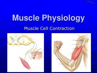
Muscle Physiology. Muscle Tissue. Muscle accounts for nearly half of the body’s mass - Muscles have the ability to change chemical energy (ATP) into mechanical energy Three types of Muscle Tissue – differ in structure, location, function, and means of activation Skeletal Muscle

Muscle Physiology
E N D
Presentation Transcript
Muscle Tissue • Muscle accounts for nearly half of the body’s mass - Muscles have the ability to change chemical energy (ATP) into mechanical energy • Three types of Muscle Tissue – differ in structure, location, function, and means of activation • Skeletal Muscle • Cardiac Muscle • Smooth Muscle
Skeletal Muscle • Skeletal muscles attach to and cover the bony skeleton • Is controlled voluntarily (i.e., by conscious control) • Contracts rapidly but tires easily • Is responsible for overall body motility • Is extremely adaptable and can exert forces ranging from a fraction of an ounce to over 70 pounds • Has obvious stripes called striations • Each muscle cell is multinucleated
Microscopic Anatomy - Skeletal Muscle Fiber • Sarcoplasm contains glycosomes (granules of glycogen) and the oxygen-binding protein called myoglobin • In addition to the typical organelles, fibers have • Sarcoplasmic reticulum • T tubules - modifications of the sarcolemma • Myofibrils • Each muscle fiber is made of many myofibrils, 80% of the muscle volume, that contain the contractile elements of skeletal muscle cells
Myofibrils - Striations • Myofibrils are made up of 2 types of contractile proteins called myofilaments • Thick (Myosin) filaments • Thin (Actin) filaments • The arrangement of myofibrils creates a series of repeating dark A (anisotropic) bands and light I (isotropic) bands
Myofibrils - Striations • The A band has a light stripe in the center called the H (helle) zone • The H zone is bisected by a dark line, the M line • I band has a darker midline called the Z disc (or Z line)
Sarcomere • Smallest contractile unit of a muscle • Myofibril region between two successive Z discs, has a central A band and partial (half) I bands at each end
Thick Filaments (16 nm diam) Myosin • Each myosin molecule (two interwoven polypeptide chains) has a rodlike tail and two globular heads • During muscle contraction, the Heads link the thick and thin filaments together, forming cross bridges
Thin Filaments - Actin • Thin filaments are mostly composed of the protein actin. • Provides active sites where myosin heads attach during contraction. Tropomyosin and Troponin are regulatory subunits bound to actin.
Sarcomere A band (c) Z disk Z disk Myofibril M line I band H zone (d) Titin Z disk Z disk M line M line Thin filaments Thick filaments Titin (f) Nebulin Troponin (e) Myosin heads Hinge region Myosin tail Tropomyosin G-actin molecule Actin chain Myosin molecule Ultrastructure of Muscle Figure 12-3c–f
Sarcoplasmic Reticulum (SR) • SR - an elaborate, smooth ER that surrounds each myofibril. Perpendicular (transverse) channels at the A band - I band junction are the Terminal Cisternae (Lateral Sacs) SR regulates intracellular Ca2+ • T tubules at each A band/I band junction - continuous with the sarcolemma. Conduct electrical impulses to the throughout cell (every sarcomere) - signals for the release of Ca2+ from adjacent terminal cisternae
Triad – 2 terminal cisternae and 1 T tubule • T tubules and SR provide tightly linked signals for muscle contraction • Interaction of integral membrane proteins (IMPs) from T tubules and SR
(b) DHP receptor SR Foot Protein (Ca++ release channel) Ca2+ Ca2+ released Interaction of T-Tubule Proteins and SR Foot Proteins • T tubule proteins (Dihydropyridine) act as voltage sensors • SR foot proteins are (ryanodine) receptors that regulate Ca2+ release from the SR cisternae • Action potential in t-tubule alters conformation of DHP receptor • DHP receptor opens Ca2+ release channels in sarcoplasmic reticulum and Ca2+ enters cytoplasm
Sliding Filament Model of Contraction • Contraction refers to the activation of myosin’s cross bridges – the sites that generate the force • In the relaxed state, actin and myosin filaments do not fully overlap • With stimulation by the nervous system, myosin heads bind to actin and pull the thin filaments • Actin filaments slide past the myosin filaments so that the actin and myosin filaments overlap to a greater degree (the actin filaments are moved toward the center of the sarcomere, Z lines become closer)
Skeletal Muscle Contraction • For contraction to occur, a skeletal muscle must: • Be stimulated by a nerve ending • Propagate an electrical current, or action potential, along its sarcolemma • Have a rise in intracellular Ca2+ levels, the final stimulus for contraction • The series of events linking the action potential to contraction is called excitation-contraction coupling
Depolarization and Generation of an AP • The sarcolemma, like other plasma membranes is polarized. There is a potential difference (voltage) across the membrane • When Ach binds to its receptors on the motor end plate, chemically (ligand) gated ion channels in the receptors open and allow Na+ and K+ to move across the membrane, resulting in a transient change in membrane potential - Depolarization • End plate potential - a local depolarization that creates and spreads an action potential across the sarcolemma
Excitation-Contraction Coupling • E-C Coupling is the sequence of events linking the transmission of an action potential along the sarcolemma to muscle contraction (the sliding of myofilaments) • The action potential lasts only 1-2 ms and ends before contraction occurs. • The period between action potential initiation and the beginning of contraction is called the latent period. • E-C coupling occurs within the latent period.
(b) Initiation of contraction Ca2+ levels increase in cytosol. 1 4 Power stroke Ca2+ binds to troponin. 2 3 Tropomyosin shifts, exposing binding site on G-actin Pi ADP Troponin-Ca2+ complex pulls tropomyosin away from G-actin binding site. 3 TN Myosin binds to actin and completes power stroke. 4 5 2 G-actin moves Actin filament moves. 5 1 Cytosolic Ca2+ Regulatory Role of Tropomyosin and Troponin Figure 12-10b, steps 1–5
Somatic motor neuron releases ACh at neuro- muscular junction. Net entry of Na+ through ACh receptor-channel initiates a muscle action potential. 1 2 (a) 1 Axon terminal of somatic motor neuron ACh Muscle fiber potential Action K+ Action potential 2 Na+ Motor end plate Sarcoplasmic reticulum T-tubule Ca2+ DHP receptor Tropomyosin Z disk Troponin Actin M line Myosin head Myosin thick filament Excitation-Contraction Coupling Figure 12-11a, steps 1–2
T tubule Terminal button Surface membrane of muscle cell Acetylcholine- gated cation channel Acetylcholine Troponin Tropomyosin Actin Cross-bridge binding Myosin cross bridge Excitation-Contraction Coupling • The action potential is propagated along (across) the sarcolemma and travels through the T tubules • At the triads, the action potential causes voltage sensitive T tubule proteins to change shape. This change, in turn, causes the SR foot proteins of the terminal cisternae to change shape, Ca2+ channels are opened and Ca2+ is released into the sarcoplasm (where the myofilaments are)
Excitation-Contraction Coupling • Some of the Ca2+ binds to troponin, troponin changes shape and causes tropomysin to move which exposes the active binding sites on actin • Myosin heads can now alternately attach and detach, pulling the actin filaments toward the center of the sarcomere (ATP hydrolysis is necessary)
Excitation-Contraction Coupling • The short calcium influx ends (30 ms after the action potential ends) and Ca2+ levels fall. An ATP-dependent Ca2+ pump is continually moving Ca2+ back into the SR. • Tropomyosin blockage of the actin binding sites is reestablished as Ca2+ levels drop. Cross bridge activity ends and relaxation occurs
Myosin filament Tight binding in the rigor state. The crossbridge is at a 45° angle relative to the filaments. ATP binds to its binding site on the myosin. Myosin then dissociates from actin. 45° Myosin binding sites 1 2 ATP binding site 3 2 4 1 G-actin molecule ADP ATP 3 2 4 1 3 2 1 4 5 6 At the end of the power stroke, the myosin head releases ADP and resumes the tightly bound rigor state. The ATPase activity of myosin hydrolyzes the ATP. ADP and Pi remain bound to myosin. 3 ADP Contraction- relaxation Pi Pi Sliding filament 3 2 4 1 3 2 4 1 5 Actin filament moves toward M line. 90° Pi 3 2 4 1 Release of Pi initiates the power stroke. The myosin head rotates on its hinge, pushing the actin filament past it. 5 The myosin head swings over and binds weakly to a new actin molecule. The crossbridge is now at 90º relative to the filaments. 4 The Molecular Basis of Contraction Figure 12-9
Motor Unit • Motor unit - One motor neuron and the muscle fibers it innervates • Number of muscle fibers varies among different motor units • Number of muscle fibers per motor unit and number of motor units per muscle vary widely • Muscles that produce precise, delicate movements contain fewer fibers per motor unit • Muscles performing powerful, coarsely controlled movement have larger number of fibers per motor unit
Electrical and Mechanical Events in Muscle Contraction A twitch is a single contraction-relaxation cycle Figure 12-12
Muscle Twitch • A muscle twitch is the response of the muscle fibers of a motor unit to a single action potential of its motor neuron. The fibers contract quickly and then relax. Three Phases: • Latent Period – the first few ms after stimulation when excitation-contraction is occurring • Period of Contraction – cross bridges are active and the muscle shortens if the tension is great enough to overcome the load • Period of Relaxation – Ca2+ is pumped back into SR and muscle tension decreases to baseline level
Graded Muscle Responses • Graded muscle responses are: • Variations in the degree or strength of muscle contraction in response to demand • Required for proper control of skeletal movement • Muscle contraction can be graded (varied) in two ways: • Changing the frequency of the stimulus • Changing the strength of the stimulus
Muscle Response to Stimulation Frequency • A single stimulus results in a single contractile response – a muscle twitch (contracts and relaxes) • More frequent stimuli increases contractile force – wave summation - muscle is already partially contracted when next stimulus arrives and contractions are summed
Muscle Response to Stimulation Frequency • More rapidly delivered stimuli result in incomplete tetanus – sustained but quivering contraction • If stimuli are given quickly enough, complete tetanus results – smooth, sustained contraction with no relaxation period
Factors Affecting Force of Muscle Contraction • Number of motor units recruited, recruitment also helps provide smooth muscle action rather than jerky movements • The relative size of the muscle fibers – the bulkier the muscle fiber (greater cross-sectional area), the greater its strength • Asynchronous recruitment of motor units -while some motor units are active others are inactive - this pattern of firing provides a brief rest for the inactive units preventing fatigue • Degree of muscle stretch
Muscle Tone • The constant, slightly contracted state of all muscles • Does not produce active movements • Keeps the muscles firm and ready to respond to stimulus • Helps stabilize joints and maintain posture • Due to spinal reflex activation of motor units in response to stretch receptors in muscles and tendons
Contraction of Skeletal Muscle Fibers • The force exerted on an object by a contracting muscle is called muscle tension, the opposing force or weight of the object to be moved is called the load. • Two types of Muscle Contraction: • When muscle tension develops, but the load is not moved (muscle does not shorten) the contraction is called Isometric • If muscle tension overcomes (moves) the load and the muscle shortens, the contraction is called Isotonic
Isometric Contractions No change in overall muscle length In isometric contractions, increasing muscle tension (force) is measured
Isotonic Contraction • In isotonic contractions, the muscle changes length and moves the load. Once sufficient tension has developed to move the load, the tension remains relatively constant through the rest of the contractile period. • Two types of isotonic contractions: • Concentric contractions – the muscle shortens and does work • Eccentric contractions – the muscle contracts as it lengthens
Isotonic Contraction This illustrates a concentric isotonic contraction In isotonic contractions, the amount of shortening (distance in mm) is measured
Energy Sources for Contraction • ATP is the only energy source that is used directly for contractile activity • As soon as available ATP is hydrolyzed (4-6 seconds), it is regenerated by three pathways: • Transfer of high-energy phosphate from creatine phosphate to ADP, first energy storehouse tapped at onset of contractile activity • Oxidative phosphorylation (citric acid cycle and electron transport system - takes place within muscle mitochondria if sufficient O2 is present • Glycolysis - supports anaerobic or high-intensity exercise
CP-ADP Reaction • Transfer of energy as a phosphate group is moved from CP to ADP – the reaction is catalyzed by the enzyme creatine kinase • Creatine phosphate + ADP → creatine + ATP • Stored ATP and CP provide energy for maximum muscle power for 10-15 seconds
Anaerobic Glycolysis • Glucose is broken down into pyruvic acide to yield 2 ATP • When oxygen demand cannot be met, pyruvic acid is converted into lactic acid • Lactic acid diffuses into the bloodstream – can be used as energy source by the liver, kidneys, and heart • Can be converted back into pyruvic acid, glucose, or glycogen by the liver
Glycolysis and Aerobic Respiration • Aerobic respiration occurs in mitochondria - requires O2 • A series of reactions breaks down glucose for high yield of ATP • Glucose + O2 → CO2 + H2O + ATP
Muscle Fatigue • Muscle fatigue – the muscle is physiologically not able to contract • Occurs when oxygen is limited and ATP production fails to keep pace with ATP use • Lactic acid accumulation and ionic imbalances may also contribute to muscle fatigue • Depletion of energy stores – glycogen • When no ATP is available, contractures (continuous contraction) may result because cross bridges are unable to detach
Muscle Fiber Type: Speed of Contraction • Speed of contraction – determined by how fast their myosin ATPases split ATP • Oxidative fibers – use aerobic pathways • Glycolytic fibers – use anaerobic glycolysis • Based on these two criteria skeletal muscles may be classified as: • Slow oxidative fibers (Type I) - contract slowly, have slow acting myosin ATPases, and are fatigue resistant • Fast oxidative fibers (Type IIA)- contract quickly, have fast myosin ATPases, and have moderate resistance to fatigue • Fast glycolytic fibers (Type IIB)- contract quickly, have fast myosin ATPases, and are easily fatigued
Smooth Muscle • Occurs within most organs • Walls of hollow visceral organs, such as the stomach • Urinary bladder • Respiratory passages • Arteries and veins • Helps substances move through internal body channels via peristalsis • No striations • Filaments do not form myofibrils • Not arranged in sarcomere pattern found in skeletal muscle • Is Involuntary • Single Nucleus
Smooth Muscle • Composed of spindle-shaped fibers with a diameter of 2-10 m and lengths of several hundred m • Cells usually arranged in sheets within muscle • Organized into two layers (longitudinal and circular) of closely apposed fibers • Have essentially the same contractile mechanisms as skeletal muscle
Smooth Muscle • Cell has three types of filaments • Thick myosin filaments • Longer than those in skeletal muscle • Thin actin filaments • Contain tropomyosin but lack troponin • Filaments of intermediate size • Do not directly participate in contraction • Form part of cytoskeletal framework that supports cell shape • Have dense bodies containing same protein found in Z lines