Ch 27: Reproductive System
This comprehensive overview explores the general organization and anatomy of the male and female reproductive systems, detailing the roles of gonads, gametes, ducts, and glands. Key structures such as the testes, epididymis, seminal vesicles, prostate gland, and penis are emphasized. The process of spermatogenesis, sperm maturation, and the significance of hormones like FSH and testosterone in reproductive health are discussed. This resource is essential for understanding reproductive anatomy and physiology.

Ch 27: Reproductive System
E N D
Presentation Transcript
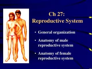
Ch 27: Reproductive System General organization Anatomy of male reproductive system Anatomy of female reproductive system
General Organization Gonads gametes & hormones Ducts transport of . . . ? Glands secrete fluid Perineal structures = external genitalia
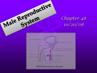
Anatomy of Male Repro System • Primary reproductive organs produce gametes • Secondary reproductive organs . . . . • Male reproductive and urinary tracts are partially shared Fig 27-1
Testes (paired glands) • Develop adjacent to kidneys • Descend into scrotum through inguinal canal • Peritoneal lining is carried along lining of scrotum • Cryptorchidism(in 3% of full-term and 30% of premature deliveries) • Significance? • Treatment? 4 month Fig 27-2
Cremaster muscle Scrotum Function: supports and protects testes Structure: Skin & underlying superficial fascia • Dartos muscle in subcutis • Cremaster muscle deep to dermis (continuation of ___________) Involuntary contraction (cremasteric reflex) in response to ________ Scrotal sac forms 2 separate chambers
Structure of Testes • Two tissue layers cover testes: • Tunica albuginea • Tunica vaginalis • 200-300 lobules • 3 seminiferous tubules Fig 27-4/5
From Spermatocyte to Spermatozoon • Spermatogenesis: Meiosis of primary spermatocytes spermatids • Spermiogenesis: Spermatid maturation into spermatozoa within Sertoli cells • Spermiation: Spermatozoon released into lumen
Sustentacular (Sertoli) Cells • Maintenance of blood testis barrier • special lumen fluid • sperm specific ag • Support of spermatogenesis • FSH and Testosterone work via Sertoli cells • Support of spermiogenesis • Secretion of inhibin • Secretion of androgen-binding protein (ABP)
Anatomy of Spermatozoon Mature sperm has 3 portions • Head with acrosome • Midpiece with lots of ? • Flagellum (rotating in corkscrew fashion) See fig 27-6
Epididymis ~ 7 m long • Sperm-maturation • Recycling of damaged spermatozoa • Adjusting composition of tubular fluid (stereocilia!!) Functions:
Path of Spermatozoa from tail of epididymis: ductus (vas) deferens ampulla ejaculatory duct urethra
Capacitation Activation of spermatozoa Occurs after spermatozoa leave epididymis and come in contact with seminal fluid. Seminal fluid + Sperm = Semen Final capacitation when exposed to conditions inside female reproductive tract
The Accessory Glands. Provide for 95% of the seminal fluid • Seminal vesicles • Prostate gland • Bulbourethral glands
Seminal Vesicles Produce 60% of seminal fluid Tubular glands (~ 15 cm) Secretion is rich in fructose leads to sperm motility
Prostate Gland • 25% of seminal fluid • Single, doughnut-shaped • Secretion contains: • citrate • seminal plasmin • prostate specific antigen (PSA)
Bulbourethral glands (Cowper’s glands) Pea size Alkaline secretion containing lots of mucus. function??
Corpora cavernosa Corpus spongiosum Erectile Tissue Penis has 3 cylindrical columns: One corpus spongiosum Two corpora cavernosa ??