Download
1 / 41
410 likes | 475 Vues
This lecture covers the types, characteristics, and functions of muscles, including skeletal muscle structure and contraction mechanisms. It explores the energetics, performance, and different types of muscle contractions. The comparison of skeletal muscle with smooth and cardiac muscle is discussed, along with the three types of muscle tissues - skeletal, cardiac, and smooth. The functions, characteristics, and structures of muscle tissues are explained in detail, including the sarcomere at the cellular and molecular levels. The mechanism of sarcomere contraction is illustrated, emphasizing the importance of ATP in the process.

E N D
Marieb’s Human Anatomy and Physiology Marieb w Hoehn Chapter 9 Muscles and Muscle Tissue Lecture 16
Lecture Overview • Types, characteristics, functions of muscle • Structure of skeletal muscle • Mechanism of skeletal muscle fiber contraction • Energetics of skeletal muscle contraction • Skeletal muscle performance • Types of skeletal muscle contractions • Comparison of skeletal muscle with smooth muscle and cardiac muscle
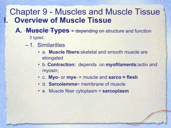
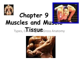
Muscular System Review - Three Types of Muscle Tissues • Skeletal Muscle • usually attached to bones • under conscious control (voluntary) • striated • multinucleated • Cardiac Muscle • wall of heart • not under conscious control • striated • branched • Smooth Muscle • walls of most viscera, blood vessels, skin • not under conscious control • not striated
Functions of Muscle • Provide stability and postural tone (skeletal) • Fixed in place without movement • Maintain posture in space • Purposeful movement (skeletal) • Perform tasks consciously, purposefully • Regulate internal organ movement and volume (mostly involuntary - smooth) • Guard entrances/exits (digestive/urinary – skeletal and smooth) • Generation of heat(thermogenesis - skeletal)
Characteristics of All Muscle Tissue • Contractile • Ability to shorten (if possible) with force; exerts tension • CANNOT forcibly lengthen • Extensible (able to be stretched) • Elastic (returns to resting length) • Excitable (can respond electrical impulses) • Conductive (transmits electrical impulses)
Structure of a Skeletal Muscle – Gross/Histological Level Figure from: Hole’s Human A&P, 12th edition, 2010 • epimysium (around muscle) • perimysium (around fascicles) • endomysium (around fibers, or cells) Alphabetical order of MUSCLE from largest to smallest: fascicle, fiber, fibril, and filament
Skeletal Muscle Fiber (Cellular level) Fully differentiated, specialized cell – its structures are given special names • sarcolemma (plasma membrane) • sarcoplasm (cytoplasm) • sarcoplasmic reticulum (ER) • transverse tubule (T-tubule) • triad • cisternae of sarcoplasmic reticulum (2) • transverse, or T-tubule • myofibril (1-2 µm diam.) Figure from: Saladin, Anatomy & Physiology, McGraw Hill, 2007 Sarcoplasmic reticulum is like the ER of other cells; but it contains [Ca2+ ] Transverse or T-tubules contain extracellular fluid ( [Na+], [K+])
Structure of the Sarcomere (Histological Level) Figures From: Marieb & Hoehn, Human Anatomy & Physiology, 9th ed., Pearson, 2013 • I band • A band • H zone • Z line • M line (~ 2µm long) The sarcomere is the contractile unit of skeletal (and cardiac) muscle
Structure of the Sarcomere (Histological/Molecular Level) ‘A’ in A band stands for Anisotropic (dArk) ‘I’ in I band stands for Isotropic (LIght) Zones of non-overlap: I band (thin filaments), and H zone (thick filaments) A sarcomere runs from Z line (disk) to Z line (disk)(From ‘Z’ to shining ‘Z’!) Figure from: Saladin, Anatomy & Physiology, McGraw Hill, 2007
Preview of Skeletal Muscle Contraction • Major steps: • Motor neuron firing • Depolarization (excitation) of muscle cell • Release of Ca2+ from sarcoplasmic reticulum • Shortening of sarcomeres • Shortening of muscle/CTs and tension produced T Tubule Sarcoplasmic reticulum Physiologyhere we come!! Figure from: Martini, Anatomy & Physiology, Prentice Hall, 2001
Grasping Physiological Concepts • The steps in a physiological process give you the ‘when’, i.e. tell you when things happen and/or the order in which they happen. • For each step in a process, you should MUST ask yourself the following questions - and be sure you get answers! • How? (How does it happen?) • Why? (Why it happens and/or why it’s important?) • What? (What happens?) See Figures 9.7 and 9.8 in your textbook for excellent overall summaries of the muscle contraction process
Sliding Filament Theory Figure from: Hole’s Human A&P, 12th edition, 2010 Theory used to explain these observations is called the sliding filament theory …
Myofilaments (Molecular Level) • Thick Filaments • composed of myosin • cross-bridges • Thin Filaments • composed of actin • associated with troponin and tropomyosin Figures From: Marieb & Hoehn, Human Anatomy & Physiology, 9th ed., Pearson, 2013
The Sarcomere as a 3D Object… https://www.youtube.com/watch?v=-pg09F5V63U
Mechanism of Sarcomere Contraction Figure from: Hole’s Human A&P, 12th edition, 2010 When you think myosin, think mover: 1. Bind2. Move3. Detach4. Reset Ca2+ troponin myosin actin
Mechanism of Sarcomere Contraction Figure from: Hole’s Human A&P, 12th edition, 2010 4. Reset 1. Bind … 3. Detach 2. Move Cycle repeats about 5 times/secEach power stroke shortens sarcomere by about 1%So, each second the sarcomere shortens by about 5% What would happen if ATP was not present? See Textbook Figure 9.12 (Focus – Cross Bridge Cycle)
Neuromuscular Junction • site where axon and muscle fiber communicate • motor neuron • motor end plate • synaptic cleft • synaptic vesicles • neurotransmitters The neurotransmitter for initiating skeletal muscle contraction is acetylcholine (ACh) Ca2+ Ca2+ SR Ca2+ Ca2+ Ca2+ Figures from: Saladin, Anatomy & Physiology, McGraw Hill, 2007
Stimulus for Contraction: Depolarization • nerve impulse causes release of acetylcholine (ACh) from synaptic vesicles • ACh binds to acetylcholine receptors on motor end plate • generates a muscle impulse • muscle impulse eventually reaches sarcoplasmic reticulum (via T tubules) and Ca2+ is released • acetylcholine is destroyed by the enzyme acetylcholinesterase (AChE) Figure from: Martini, Anatomy & Physiology, Prentice Hall, 2001 Linking of nerve stimulation with muscle contraction is called excitation-contraction coupling (See Fig 9.11 in textbook)
Summary of Skeletal Muscle Contraction Figure from: Martini, Anatomy & Physiology, Prentice Hall, 2001 5. Contraction Cycle begins - Bind (Ca, myosin) - Move - Detach - Reset Contraction Relaxation See Textbook Figure 9.12 (Focus – Cross Bridge Cycle)
Modes of ATP Synthesis During Exercise Muscle stores enough ATP for about 4-6 seconds worth of contraction, but is the only energy source used directly by muscle. So, how is energy provided for prolonged contraction? Continual shift from one energy source to another rather than an abrupt change Figures From: Marieb & Hoehn, Human Anatomy & Physiology, 9th ed., Pearson, 2013
Energy Sources for Contraction (Creatine-P) Figures From: Marieb & Hoehn, Human Anatomy & Physiology, 9th ed., Pearson, 2013 myoglobin stores extra oxygen so it can rapidly supply muscle when needed
Oxygen Debt (Excess Post Exercise O2Consumption – EPOC) EPOC - amount of extra oxygen needed by liver to convert lactic acid to glucose, resynthesize creatine-P, make new glycogen, and replace O2 removed from myoglobin. • when oxygen is not available • glycolysis continues • pyruvic acid converted to lactic acid (WHY?) • liver converts lactic acid to glucose Figure from: Hole’s Human A&P, 12th edition, 2010 (The Cori Cycle)
Muscle Fatigue • Inability to maintain force of contraction although muscle is receiving stimulus to contract • Commonly caused by • decreased blood flow • ion imbalances • accumulation of lactic acid • relative (not total) decrease in ATP availability • decrease in stored ACh • Cramp – sustained, involuntary contraction
Length-Tension Relationship Figures From: Marieb & Hoehn, Human Anatomy & Physiology, 9th ed., Pearson, 2013 Maximum tension in striated muscle can only be generated when there is optimal (80-100%) overlap between myosin and actin filaments
Muscular Responses • Threshold Stimulus • minimal strength required to cause contraction in an isolated muscle fiber Figure From: Marieb & Hoehn, Human Anatomy & Physiology, 9th ed., Pearson, 2013 • Record of a Muscle Contraction = myogram • latent period • period of contraction • period of relaxation • refractory period • all-or-none response An individual muscle fiber (cell) is either “on” or “off” and produces maximum tension at that resting length for a given frequency of stimulation
Treppe, Wave Summation, and Tetanus • Treppe, Wave Summation, and Tetanus • all involve increases in tension generated in a muscle fiber after more frequent re-stimulation • The difference among them is WHEN the muscle fiber receives the second, and subsequent, stimulations: • Treppe – stimulation immediately AFTER a muscle cell has relaxed completely. • Wave Summation – Stimulation BEFORE a muscle fiber is relaxed completely • Incomplete (unfused) tetanus – partial relaxation between stimuli • Complete (fused) tetanus – NO relaxation between stimuli
Treppe, Wave Summation, and Tetanus Wave (Temporal) Summation Treppe (10-20/sec) Little/no relaxation period Complete Tetanus (>50/sec) Incomplete Tetanus (20-30/sec) Tetany is a sustained contraction of skeletal muscle Figure from: Martini, Anatomy & Physiology, Prentice Hall, 2001
Motor Unit • single motor neuron plus all muscle fibers controlled by that motor neuron Figure From: Marieb & Hoehn, Human Anatomy & Physiology, 9th ed., Pearson, 2013
Recruitment of Motor Units • recruitment - increase in the number of motor units activated to perform a task • whole muscle composed of many motor units • as intensity of stimulation increases, recruitment of motor units continues, from smallest to largest, until all motor units are activated
Sustained Contractions • smaller motor units recruited first • larger motor units recruited later • produces smooth movements • muscle tone – continuous state of partial contraction
Types of Contractions • concentric– shortening contraction • isotonic – muscle contracts and changes length • eccentric – lengthening contraction • isometric – muscle “contracts” but does not change length Figure from: Hole’s Human A&P, 12th edition, 2010
Types of Skeletal Muscle Fibers All fibers in any given motor unit are of the same type
Types of Skeletal Muscle Fibers All fibers in any given motor unit are of the same type
Smooth Muscle Fibers • Compared to skeletal muscle fibers • shorter • single nucleus • elongated with tapering ends • myofilaments organized differently • no sarcomeres, so no striations • lack transverse tubules • sarcoplasmic reticula not well developed • exhibit stress-relaxation response (adapt to new stretch state and relax) Figure from: Martini, Anatomy & Physiology, Prentice Hall, 2001
Types of Smooth Muscle • Single-unit (unitary) smooth muscle • visceral smooth muscle • sheets of muscle fibers that function as a group, i.e., a single unit • fibers held together by gap junctions • exhibit rhythmicity • exhibit peristalsis • walls of most hollow organs, blood vessels, respiratory/urinary/ reproductive tracts • Multiunit Smooth Muscle • fibers function separately, i.e., as multiple independent units • muscles of eye, piloerector muscles, walls of large blood vessels
Smooth Muscle Contraction • Resembles skeletal muscle contraction • interaction between actin and myosin • both use calcium and ATP • both depend on impulses • Different from skeletal muscle contraction • smooth muscle lacks troponin • smooth muscle depends on calmodulin • two neurotransmitters affect smooth muscle • acetylcholine and norepinephrine • hormones affect smooth muscle • have gap junctions • stretching can trigger smooth muscle contraction (but briefly, then relaxation again occurs) • smooth muscle slower to contract and relax • smooth muscle more resistant to fatigue • smooth muscle can undergo hyperplasia, e.g., uterus
Cardiac Muscle • only in the heart • muscle fibers joined together by intercalated discs • fibers branch • network of fibers contracts as a unit (gap junctions) • self-exciting and rhythmic • longer refractory period than skeletal muscle (slower contract.) • cannot be tetanized • fatigue resistant • has sarcomeres Figure from: Martini, Anatomy & Physiology, Prentice Hall, 2001
Review • Three types of muscle tissue • Skeletal • Cardiac • Smooth • Muscle tissue is… • Contractile • Extensible • Elastic • Conductive • Excitable
Review • Functions of muscle tissue • Provide stability and postural tone • Purposeful movement • Regulate internal organ movement and volume • Guard entrances/exits • Generation of heat • Muscle fiber anatomy • Actin filaments, tropomyosin, troponin • Myosin filaments • Sarcomere • Bands and zones
Review • Muscle contraction • Sliding filament theory • Contraction cycle (Bind, Move, Detach, Release) • Role of ATP, creatine • Metabolic requirements of skeletal muscle • Stimulation at neuromuscular junction • Muscular responses • Threshold stimulus • Twitch – latent period, refractory period • All or none response • Treppe, Wave summation, and tetanus
Review • Muscular responses • Recruitment • Muscle tone • Types of muscle contractions • Isometric • Isotonic • Concentric • Eccentric • Fast and slow twitch muscle fibers • Slow Oxidative (Type I) (think: REDSOX) • Fast Oxidative-glycolytic (Type II-A) • Fast Glycolytic (Type II-B)