Download
1 / 72
720 likes | 2.04k Vues
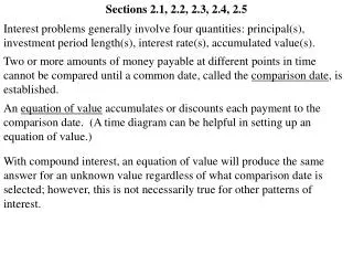
Abdomen 2. 2.3 Peritoneal cavity 2.4 Liver and gallbladder 2.5 Stomach and spleen. Albert van Schoor GNK 288 (SA4 Anatomy dissection). 2.3 Peritoneal cavity and Disposition of abdominal organs. 2.3.1 Peritoneum 2.3.2 Organs and relations 2.3.3 Peritoneal specialisation

E N D
Abdomen 2 2.3 Peritoneal cavity2.4 Liver and gallbladder2.5 Stomach and spleen Albert van Schoor GNK 288 (SA4 Anatomy dissection)
2.3 Peritoneal cavityand Disposition of abdominal organs 2.3.1 Peritoneum 2.3.2 Organs and relations 2.3.3 Peritoneal specialisation 2.3.4 Radiographic anatomy
2.3.1 Peritoneum • Define the terms: • Peritoneum, • Omentumand • Mesentery • Identify the parietal and visceral peritoneum • Identify and briefly discuss the attachments of the greater and lesser omentum
2.3.1 Peritoneum • Serous membrane that lines the abdominopelvic cavity and invests the viscera • Parietal peritoneum • Visceral peritoneum • Intraperitoneal organs • Stomach, spleen • Retroperitoneal organs • Kidneys, ascending & descending colon
2.3.1 Peritoneum Mesentry of jejenum and ileum Transverse mesoclolon Sigmoid mesocolon Meso-appendix
Lesser Omentum 2.3.1 Peritoneum Greater Omentum
2.3.1 Peritoneum • Innervation • Central aspect, diaphragmatic peritoneum • Phrenic nerve (C3-5) (referred pain) • Peripheral aspect, diaphragmatic peritoneum • Inter- and subcostal nerves (T7-T12) • Parietal peritoneum • T7-T12 and L1 (pain at precise point of stimulation) • Visceral peritoneum • Insensitive to mechanical stimulation
2.3.2 Organs and relations • Explain the functional anatomy of the mesentery, it’s position, vascular, lymphatic and neural contents • Explain how the abdomen is divided into a supracolic and infracolic compartment • Identify and briefly discuss the attachments of the mesentery of the small intestine to divide the infracolic compartment in two regions
2.3.2 Organs & relations • Mesentry: • double layer of peritoneum • serves as continuation of visceral and parietal peritoneum • provides a means for neurovascular communication between organ and body wall
2.3.2 Organs & relations Transverse Mesoclolon Mesentry of jejenum and ileum Sigmoid mesocolon
2.3.2 Organs & relations Supracolic Infracolic Right Left
2.3.3 Peritoneal specialisation • Name and identify the peritoneal folds • Name and identify the peritoneal fossae • Name and identify the paracolic gutters
2.3.3 Peritoneal specialisation Folds Inferior to the umbilicus • Reflection of peritoneum • Raised from abdominal wall by underlying structure • Median umbilical fold – urachus • Medial umbilical fold – obliterated umbilical artery • Lateral umbilical fold – inferior epigastric vessels
2.3.3 Peritoneal specialisation • Folds • Superior to the umbilicus • Falciform ligament • Round ligament of the liver (obliterated foetal umbilical vein)
2.3.3 Peritoneal specialisation Fossae / Recess • Duodenal recess • Caecal recesses: • Superior ileocaecal • Inferior ileocaecal • Retrocaecal • Intersigmoid recess • Omental bursa
2.3.3 Peritoneal specialisation • Fossae / Recess • Duodenal recess • Duodenojejunal flexure • Formed by superior and inferior duodenal folds • Superior and inferior duodenal recesses • Paraduodenal recess
2.3.3 Peritoneal specialisation • Fossae / Recess • Caecal recesses: • Superior ileocaecal • Inferior ileocaecal • Retrocaecal • Formed by: • Caecal fold • Ileocaecal fold • Vascular fold
2.3.3 Peritoneal specialisation • Fossae / Recess • Caecal recesses: • Superior ileocaecal • Inferior ileocaecal • Retrocaecal • Formed by: • Caecal fold • Ileocaecal fold • Vascular fold
2.3.3 Peritoneal specialisation • Fossae / Recess • Intersigmoid recess • Meso-sigmoid attached to posterior abdominal wall in relation where the left ureter crosses the left common iliac artery
2.3.3 Peritoneal specialisation Gutters • Right paracolic gutter • Left paracolic gutter • Right, between mesentry of jejenum and ileum and ascending colon (no exit) • Left, between mesentry of jejenum and ileum and descending colon (exit inferior)
2.3.3 Peritoneal specialisation • Identify the following: • Gastrosplenic ligament, • Splenorenalligament, • Transverse and sigmoid mesocolon, • Ileocoecal fold, • Meso-appendix and • The mesenterium of the small intestine
2.3.3 Peritoneal specialisation • Identify and describe the omental bursa (lesser sac) in respect of its relations, borders and entrance - the omental foramen • Identify the structures forming the borders of the omental foramen • Name and identify the subphrenic spaces
2.3.3 Peritoneal specialisation • Omental bursa • (left subhepatic space) • Superior recess • Inferior recess • Splenic recess
2.3.3 Peritoneal specialisation • Omental bursa • Superior recess • Anterior: • Lesser omentum • Caudate process of liver • Posterior: • Diaphragm • Right: • IVC • Left: • Oesophagus
2.3.3 Peritoneal specialisation • Omental bursa • Inferior recess • Anterior: • Stomach • Anterior 2 layers of greater omentum • Posterior: • Pancreas, transverse colon and mesocolon, poster 2 layers of greater omentum
2.3.3 Peritoneal specialisation • Omental bursa • Splenic recess • Anterior: • Gastrosplenic ligament • Posterior: • Splenorenal ligament • Left: • Hilum of the spleen
2.3.3 Peritoneal specialisation • Omental foramen • Connects greater sac (peritoneal cavity) and lesser sac (omental bursa)
2.3.3 Peritoneal specialisation • Omental foramen • Anterior: • Free border of lesser omentum: • Common bile duct (right) • Proper hepatic artery (left) • Hepatic portal vein (posterior)
2.3.3 Peritoneal specialisation • Omental foramen • Posterior: • Inferior vena cava
2.3.3 Peritoneal specialisation • Omental foramen • Superior: • Caudate lobe of liver
2.3.3 Peritoneal specialisation • Omental foramen • Inferior: • 1st part of duodenum • Common hepatic artery
2.3.3 Peritoneal specialisation • Name and identify the subphrenic spaces
2.3.3 Peritoneal specialisation • Spaces • Right and left subphrenic spaces (separated by the falciform ligament) • Right subhepatic space (Morison’s pouch) • Left subhepatic space (omental bursa) • Extraperitoneal subphrenic space
2.3.3 Peritoneal specialisation • Right and left subphrenic spaces (separated by the falciform ligament)
2.3.3 Peritoneal specialisation • Right subhepatic space (Morison’s pouch)
2.3.3 Peritoneal specialisation • Extraperitoneal subphrenic space
2.3.4 Radiographic anatomy • Identify the following structures on a plain erect abdominal X-ray: • ASIS, • lumbar vertebrae, • SI-joint, • large intestine, • diaphragm, • stomach with air in fundus of stomach, • liver, • psoas line www.up.ac.za/academic/medicine/anatomy/current/sa4/week01e.html#radio
2.4 Liver and gallbladder 2.4.1 Surface anatomy 2.4.2 Structure 2.4.3 Blood supply, nerve supply and lymph drainage
2.4.1 Surface anatomy • Review the surface anatomy of the liver and gallbladder • Indicate where a liver biopsy should be done
2.4.1 Surface anatomy • Liver • Right, midaxillary line: • 7th rib almost to right iliac crest • Right, midclavicular plane: • 5th rib cartilage to 9th costal cartilage • Left, midclavicular plane: • 2.5cm short 5th intercostal space and left nipple
2.4.1 Surface anatomy • Gallbladder • Inferior to 9th costal cartilage on right • Lateral to semilunar line (lateral border of rectus abdominis) • Approx. hands breadth from midline
2.4.2 Structure • Name and identify the borders and surfaces of the liver • Name and identify the lobes, segments, fissures with their contents Identify the subhepatic and subphrenic spaces, and their possible implication in the spread of infection
2.4.2 Structure Diaphragmatic surface Visceral surface
2.4.2 Structure Left Right
2.4.2 Structure • Colon • Kidney • Duodenum • Stomach
2.4.2 Structure • Name and identify the following: • Triangular ligaments, • Coronary ligaments, • Falciform ligament, • Lesser omentum, • Round ligament of the liver and • Ligamentum venosum
2.4.2 Structure • Identify, schematically illustrate and discuss the extrahepatic bile ducts as follows: • Origin, • Course, • Outlet and • Relations to the pancreas head and duodenum • Name and identify the extrahepatic bile ducts on radiographs
2.4.2 Structure Right and left hepatic ducts Common hepatic duct Cystic duct (Common) bile duct Main pancreatic duct of Wirsung Ampulla of Vater with the sphincter of Oddi
ERCPEndoscopic retrograde cholangiopancreatography 2.4.2 Structure