Download
1 / 39
390 likes | 404 Vues
Explore advanced microscopy techniques and enhancement methods for 3D reconstruction in nano-scale imaging. This project involves revising and implementing improvements based on previous work by others.

E N D
Publication Project and Microscopy Review Imaging, Robotics, & Intelligent Systems Laboratory University of Tennessee Chris Kammerud Summer 2004
Outline • Instrumentation/Publication Project • Review other’s previous work • Revisements to paper • Implementation • Reviewed Matt’s previous work • Collected Information on microscopes in SERF Lab • Collected data with Matt • Survey of Nano-scale Computer Vision • Enhancment methods • 3D reconstruction systems • Survey of Nano-scale Instruments • NSOM/SNOM • AFM • Conclusions and Future Work
Instrumentation/Publication Objectives • Continue my previous work luggage simulation algorithm and Viz-Tek • Revise report based on suggestions and with more references to previous work
Others Work in Luggage Simulation • Alstec: Airport Technology – Baggage Systems Simulation Tests. [Als04] • Airport Luggage Inspection Simulation with Force Feedback. [Har04] • EDTECH AIR Security Simulator. [Edt03] • Explosives detection systems (EDS) for aviation security. [Sin03] • Innovations, Inc. TIP – Threat Image Projection. [TIP04] • Research Projects – Airport Luggage Handling, Airport simulation library. [Val04]
Revising paper • Included more references to work mentioned on previous slide • Focus on contributions and differences with work I did compared to the work of others • Much work was done, ([Als04] and [Val04] ) with high level simulation, i.e. simulating the flow of passengers through security with models of luggage screening. My research is at a lower level, building simulated models of packed luggage • Work has also been done in simulating x-rays of luggage ([TIP04] and [Edt03] ). My research was in developing virtual models of luggage and does not use or need previous x-rays of real luggage
Results • A revised paper was created with new sections on motivation, contribution, and a few others. • Previous work was researched and read to determine the contribution of my work
Plan for Inst/Pub Project • Further changes will be made according to the suggestions received in the past days • Inquiries into when and if Viz-Tek system can be reassembled • Depending on amount of work needed to be done with report, refinements could be made to the luggage simulation script in 3DSM.
Implementation Objectives • Familiarize self with microscopes in SERF • Review Matt and Sherry’s work • Collect own data • Expand on data by mosaicing
Review of Matt and Sherry’s work • Matt’s work looked at includes • “The State of the Art of Range Imaging” [Sch02] • “Image Acquisition and 3D Model Reconstruction of Biomedical Specimens” [Sch03] • “SEM Preparation” section of master thesis paper he’s working on • Sherry’s work looked at • “Microscopic Scanning” [Ali04] • Got book on Microfabrication from her
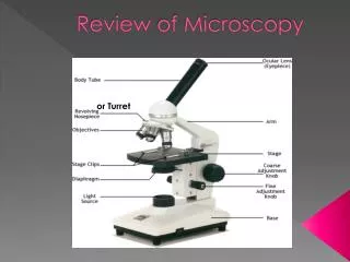
Familiarization with Microscopes • Went with Matt to learn SEM • Learned how to prepare samples • Used SEM to collect images
Microscopy Implementation Plan • Matt is going down for a second scan to take images of calibration spheres with SEM, I will go with him and collect more data • Specifically collect data for the process of mosaicing • Implement mosaicing
Survey of Nano-Scale CV Methods Objectives • Review of algorithms for nano-scale devices • Review methods and systems for 3D reconstruction
Enhancement algorithms • “Noise adaptive channel smoothing for low-dose images” [Sch et al03] • “Enhanced feature analysis using wavelets for scanning probe microscopy images of surfaces” [Mak et al04]
Noise adaptive channel smoothing • Nano-scale images have different noise characteristics than camera based, Poisson vs Gaussian • Channel smoothing • Each pixel value has N vector weight(N channels) attached to it • Channels are smoothed individually • Image is reconstructed from smoothed channels
Channel Smoothing cont • Adapting to Poisson • Low-intensity images with Poisson noise, the noise variance increases at increased gray-level values • Smoothing is done per channel with Gaussian function, higher channels(higher gray-levels) are smoothed with Gaussian of higher variance Channel smoothing information and images from [Sch et al03]
Wavelet Analysis on AFM • Uses wavelets to denoise 3D measurement data [Mak et al04]
3D Reconstructions • “3D Modeling from AFM measurements” [JosHug01] • “High Resolution 3D X-ray Diffraction Microscopy” [Jia et al02] • “Innovative Metrology Method for the 3-Dimensional Measurements of MEMS” [Wol et al04]
3D Modeling from AFM • Similar to range scanning of models we do at IRIS • AFM measurements are taken with the sample at different orientations to the scanner • These models are combine into a more complete 3D model
3D Modeling from AFM, Block Diagram Block diagram of system proposed, [JosHug01]
3D Modeling from AFM (Results) Two views of quartz particle model combined [JosHug01]
3D X-ray Diffraction Microscopy • Microscopy combining coherent x-ray diffraction with the oversampling phasing method • X-rays probe deeper into specimen than electrons but are harder to focus • Resolution from this paper’s method is 8 nm for 2D image and 50 nm for the 3D reconstruction [Jia et al02]
3D X-ray Diffraction Microscopy, 2D results Electron scan X-ray scan, lower layer information visible [Jia et al02]
3D X-ray Diffraction Microscopy, 3D results [Jia et al02]
3-D of MEMS • Phase Shift Inteferometry • Known wavefront interfering with unknown waves reflected from specimen • Paper presents method without moving mirror • Measurement of certified step hight within 0.4 nm [Wol et al04]
3-D of MEMS, Results Silicon Pit Cantilever [Wol et al04]
Plans on CV Survey • Continue looking at papers and new research • Write into report as collect
Survey of Nano-Scale Instrument Objectives • Collect information on commercially available products • Microscopes • Display systems, accessories, etc.
Commercial Microscopes • Types of Microscopes • Standard Optical Light • Confocal • TEM • SEM • AFM • NSOM/SNOM • The bottom 4 are best able to image with resolutions < 100 nm • Predominant time was spent on NSOM/SNOM, since previous work by Matt and Sherry had not discussed this technology
TEM • In SERF Lab • Hitachi H-800 • 200 kV operating voltage • Resolution 0.3 nm Hitachi H-800 TEM image of magnetoreistance spin valve multilayer [MSU03]
SEM • Available in SERF • Leo 1525 • 1.5 nm resolution at 20 kV and 3.5 nm at 1 kV [Leo04]
AFM • Recently added to SERF • Asylum MFP-3D • Noise level in Z < 0.2 nm, and < 0.4 nm for X and Y directions [Asy04]
NSOM/SNOM • SPM technology, light passed through tiny aperture • Four modes, transmission, reflection, collection, illumination [NSO04]
Nanonics Image 30 nm gold balls Nanonics NSOM in upright microscope Images from http://www.nanonics.co.il/main/prod_item.php?ln=en&item_id=80&cat=5&sub_cat=22
Plan for Instruments • Focus on systems such as AFM, SEM • Research stages, cameras, etc.
Conclusions and Future Work • 4 tasks were presented • Instrumentation/Publication • Implementation with microscopes • Review of nano-scale computer vision methods • Review of nano-scale instrumentation • Work will continue on each task as stated in previous sections • References are on the following slides
References [Ali04] Ali, S. “Microscopic Scanning”, http://imaging.utk.edu/classes/spring2004/ngozi/599/webfiles/reports.htm, 2004. [Asy04] Asylum Research MFP-3D http://www.af-fe.de/Asylum_Research/asylum_research.html, 2004. [Als04] Alstec: Airport Technology – Baggage Systems Simulation Tests. http://www.airport-technology.com/contractors/baggage/alstec/alstec3.html, 2004. [Edt03] J. Leibow, L. Gomez, L. Clemente: EDTECH AIR Security Simulator. http://eprentice.sdsu.edu/F03670/securitysim/, 2003. [Har04] E. Harper: Airport Luggage Inspection Simulation with Froce Feedback.http://www.caip.rutgers.edu/vrlab/projects/luggage/, 2004. [Jia et al02] M. Jianwei, T. Ishikawa, B. Johnson, E.H. Anderson, B. Lai, K.O. Hodgson, “High Resolution 3D X-ray Diffraction Microscopoy.” Physical Review Letters, 19 Aug 2002. [JosHug01] T. Jost, H. Hugli, “3D modeling from AFM measurments.” Proceedings of the SPIE The International Society for Optical Engineering, 4275, p 61-70, 2001.
References cont. [Leo04] LEO 1525 Field Emission Scanning Electron Microscope. http://www.ubicampus.mh-hannover.de/~physik/materialsammlung/med12/Leo_sem1525.htm. 2004. [Mak et al04] A. Maksumov, R. Vidu, A. Palzoglu, P. Stroeve, “Enhanced feature analysis using wavelets for scanning probe microscopy images of surfaces”, Journal of Colloid and Interface Science, vol 272, issue 2, p 365-377, 2004. [MSU03] “Analytical Characterization Facilities: Hitachi H-800 Transmission Electron Microscope”, http://www.chems.msu.edu/resources/hitachih800.htm, 2003. [NFS04] “Near-Field Scanning Optical Microscopy”, http://www.olympusmicro.com/primer/techniques/nearfield/nearfieldintro.html, 2004 [Sch et al03] H. Scharr, M. Felsberg, P.E. Forsen, “Noise adaptive channel smoothing of low-dose images.” Workshop Computer Vision for the Nano-Scale, CVPR2003, 2003. [Sch02] M. Schultz, “State of the Art of Range Imaging as of 2002”, http://imaging.utk.edu/research/schultz/presentations.htm, 2002. [Sch03] M. Schultz, “Image Acquistion and 3D Model Reconstruction of Biomedical specimens”, http://imaging.utk.edu/research/schultz/presentations.htm, 2003.
References cont. [Sin03] S. Singh, M. Singh: Explosives detection systems (EDS) for aviation security. Signal Processing 83, 31-55, 2003. [TIP04] American Innovations, Inc. TIP – Threat Image Projection.http://www.bombdetection.com/tip.shtml, 2004. [Val04] E. Valentin: Research Projects – Airport Luggage Handling, Airport simulation library. http://www.tbm.tudelft.nl/webstaf/edwinv/, 2004. [Wol et al04] S. Wolfling, D. Banitt, Y.N., Ben, Y. Arieli, “Innovative Metrology Method for the 3-Dimensional Measurements of MEMS.” Proceedings of SPIE The International Society for Optical Engineering, v 5343, p 255- 263, 2004.