Excitable Tissues- Nerve
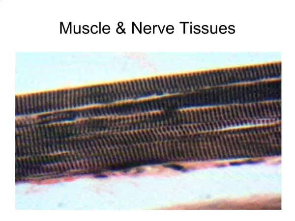
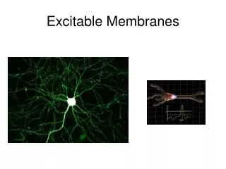
Excitable Tissues- Nerve. Prof. K. Sivapalan. Neuron. There are many types of cells. Neuron is the main functional cell in the nervous system. Cell body and processes: axon and dendrites


Excitable Tissues- Nerve
E N D
Presentation Transcript
Excitable Tissues- Nerve Prof. K. Sivapalan
Neuron • There are many types of cells. • Neuron is the main functional cell in the nervous system. • Cell body and processes: axon and dendrites • Axons transmit impulses from the cell body, covered by Schwan cells- Myalinsheeth and Nodes of Ranvier • Cutaneous nerves- exception Nerve
Membrane Potential • When two electrodes are placed, one inside and the other outside, a potential difference is observed. • It is ‘resting membrane potential’ Nerve
Basis of Resting Membrane Potential:Gibbs–Donnan effect • Cell membrane does not conduct electricity • Both sides of the membrane contains cations and anions • Based on the permiability of the individual ions, imbalance of electrical and chemical neutrality is generated Nerve
Physics of Membrane Potential • Diffusion potential is generated by ion concentration difference on both sides of the membrane • Nernst equation: Nerve
EMF with Many Ions • Goldman-Hodgkin-Katz equation: • C- Concentration, P-Permiability Nerve
Genesis of Resting Membrane Potential • Potassium diffusion potential: concentration ratio is 35:1 contributing to -94 mV • Sodium diffusion potential: concentration ratio is 0.1, contributing to +61 mV [permeability is 100 times less reducing the contribution] • Sodium Potassium pump: [3 Na+:2K+] contributes for -4 mV. [Energy] Nerve
Change in Resting Membrane Potential • The resting membrane potential can be disturbed by external electrodes or mechanical / chemical factors that alter membrane permeability. • If the potential changes towards zero, it is hypo polarization and if it becomes more negative, it is hyper polarization- Local response. • This change is proportional to the strength of the stimulus Nerve
Local Response Nerve
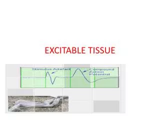
Action Potential • When the local response goes above -55 mV [firing or threshold level], the potential automatically goes to +35 [Depolarization] and immediately returns to the resting value [Repolarization] • It is known as ‘Action Potential’ Nerve
Ionic Basis of Action Potential • Membranes of nerves has voltage gated sodium and potassium channels. • When the threshold level is reached, voltage gated sodium channels open and close almost immediately. • As the channels open, sodium ions rush inwards taking positive charge inwards reversing the membrane potential- Depolarization. • Very soon voltage gated potassium channels open allowing the potassium ions out resulting in positive charge going out – Repolarization • Sodium – Potassium (ATP ase) pump and diffusion of Potassium ions bring the ionic composition back to normal Nerve
Components of Action Potential • Resting membrane potential • Firing level • Depolarization • Repolarization • After hyperpolarization • Refractory period • Energy requirements Nerve
Spread of Action Potential • Local response affects only the area stimulated. • When the membrane has an area with +ive charge outside and –ive inside, it sets up flow of current which alters the potential difference of the adjacent area • When it is due to action potential , it is sufficient to bring the potential to the threshold level and action potential is generated. • A wave of depolarization spreads across the membrane by auto stimulation of the adjacent area followed by repolarization Nerve
Conduction in Myelinated Nerve • The myelin sheeth is an insulator and it does not permit exchange of ions across the membrane. • The nodes of Ranvier contains exposed membrane with voltage gated channels for sodium and potassium. • When one node of Ranvier is depolarized, it stimulates the next node. • This results in action potentials jumping from one node to the next by passing the membrane covered by myelin [saltatory conduction]. • When compared to non-myelinated nerve, myelinated nerve conducts the impulse faster Nerve
Myelination • The Schwann cell envelops the axon and the cell raps round several times. • Each cell is 1-3 mm in length • The exposed axon in between is about 2-3 • Energy need- less Nerve
Velocity of Conduction • Myelination- faster • Larger diameter- faster • Range- 0.25 to 100 M/Sec Nerve
Susceptibility of Nerves Nerve