Download
1 / 1
10 likes | 161 Vues
Activated Eosinophils Interacting with Periostin, a Protein Found in Asthma Manav Khanna, Mats Johansson, Deane Mosher Department of Biomolecular Chemistry, University of Wisconsin-Madison. Abstract. Methods. Results.

E N D
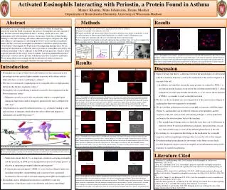
Activated Eosinophils Interacting with Periostin, a Protein Found in Asthma Manav Khanna, Mats Johansson, Deane Mosher Department of Biomolecular Chemistry, University of Wisconsin-Madison Abstract Methods Results Eosinophils are a type of leukocytes that contribute to asthma by migrating selectively from the blood-stream into the airways. Eosinophils can only migrate if they become activated and polarized, that is, develop a front and a rear. Cell polarization starts with mediator proteins, for instance, from the cytokine family, binding to cells and activating cell surface adhesion receptors, integrins, that help cells adhere to and migrate on cells lining the blood vessels and the extracellular matrix (ECM). Activated eosinophils form adhesive structures called podosomes (“foot bodies”) that degrade ECM proteins when migrating through them. We are studying the distributions of different surface proteins in eosinophils activated by the cytokine interleukin-5 (IL-5) adherent to the ECM protein periostin, which is found in greater amounts in the airway tissue in asthma. I am using immunofluorescence microscopy to detect surface proteins with fluorescent dyes coupled to antibodies. We are using fluorescent staining and the localization of surface proteins as molecular markers for eosinophils that have become activated. I Wells with coverslips were coated with 5 μg/ml periostin substrate solution II Purified eosinophils were added to each well III Primary antibodies and fluorescently labeled secondary antibodies were added, sequentially, to stain for surface proteins or fluorescently labeled phalloidin was added to stain for filamentous actin IV Coverslips were mounted and cells were viewed using fluorescent microscopy We have conducted 8 experiments in all and experiments take anywhere from 1-2 weeks to complete the assay and photo analysis. Majority of the time, cells were viewed with 100X objective. A A III I (Immunomount) II Microscope Camera Figure 5: Eosinophils treated with IL-5 and adherent on periostin viewed under 60x magnification. The cells are stained for PSGL-1 and were viewed using confocal microscopy. Both images (A-B) show the fourth layer from the bottom of thirteen layers. Image A shows three different two dimensional planes with the focus on the same cell (indicated by the white arrows): image I shows the XY plane, image II shows the XZ plane, and image III show the YZ plane. Image B shows a three dimensional view of al layers of the same sample. The height of each layer is 0.5μm. I & II III IV IV Results Introduction Discussion +IL-5 –IL5 • Eosinophils are a type of white blood cells (leukocytes) that are present in low percentages in every person, higher numbers in persons with asthma, and are believed to have a role in contributing to asthma2,3 • The true or evolutionarily original purpose of eosinophil cells is to fight parasitic infections that the host organisms contract1 • Eosinophils’ role in contributing to asthma is caused by their migration from the blood stream into airway3 • Migration can only occur if cells are polarized, which is a morphological change in shape from round to elongated; polarized cells have a defined front and a rear2 • Cell polarization is caused by mediator proteins, e.g., cytokines, binding to cells and activation of integrins, which allow the cells to adhere and migrate on endothelial cells and ECM proteins2,3,6 • Studies have shown that IL-5 is an important cytokine in activating eosinophils and that periostin, an ECM protein upregulated in airways of asthma patients, is effective in supporting eosinophil adhesion and migration3 • P-selectin glycoprotein ligand-1 (PSGL-1) is involved in rolling of leukocytes, including eosinophils, on endothelium and is known to have a polarized localization at the rear end of activated migrating neutrophils and lymphocytes6 • Eosinophilic airway inflammation is known to contribute to some key characteristics of the disease such as exacerbations and airway remodeling4 • Figure 2 reveals that there is a difference between the morphologies of cells treated with IL-5 and those that aren’t, such as the localization of the nucleus (Figure 4) at one end of the cell • In addition, we found that, among the many proteins we stained for, PSGL-1 is one surface protein localizes at one end of the cell when treated with IL-5, albeit it might not be at the same end that the nucleus is; so we can use the localization of PSGL-1 as a marker to study eosinophil activation • We also see that eosinophils can cause degradation of the periostin layer (Figure 4) implying that there was migration by eosinophils • We also find that polarization can cause eosinophils to form into a hill-like shape (Figure 3), and that there can be different structures at the periphery and the “summit” of the cell, such as in the actin staining in Figure 3, where podosomes are found in the substrate plane, but not the top plane • This morphological change makes us think that since there can be differences in structure viewed by moving to different focal planes, that in the future, we can use confocal microscopy to view all the different planes/layers of the cells • By studying we can expand our knowledge on the mechanism for eosinophil migration and the morphological changes that occur in the cells as they migrate • By understanding the mechanism for movement, in the future we may find a possible therapeutic target to prevent eosinophil caused inflammation and possibly learn how to control exacerbations PSGL-1 D C B A Actin F E Figure 2: Eosinophil cells adherent on periostin. The top row (A-D) shows cells stained for p-selectin glycoprotein ligand-1: cells treated with IL-5 (A-B) show localization of PSGL-1at one end of the cell indicating cell polarization (and cells were more spread out). Some cells after IL-5 treatment showed PSGL-1 localization in the same end as the nucleus (B). Cells not treated with IL-5 (C-D) show no polarization, but have PSGL-1 around entire border of the cell and are rounder in shape. Cells in the bottom row (E-F) are stained for actin, which is punctate in some cells. Cells treated with IL-5 (E) show localization of the nucleus at one end indicating polarization, whereas, in untreated cells, (F), actin is around the cell border and are more round in shape. Substrate Plane Higher Plane Phase PSGL-1 A B C Actin E D F Figure 3: Eosinophil cells treated with IL-5 and adherent on perisotin. The fluorescent images in each row show the same cells, just viewed at different focal planes. Phase contrast images (C, F) taken was viewed at same focal plane as “Higher Plane” images. Images A and D show the substrate focal plane, and this view reveals the actual periphery of the cells. At this plane, one can see podosomes on the cells stained for actin. These images reveal that not only do eosinophils become polarized if treated with IL-5, they can also develop a hill-like shape. At a higher focal plane (B, E) only the “summit” of the cell is seen, and a more clear image of PSGL-1 localization (B) is also seen. Evans R et al. J Cell Sci 2009;122:215-225 Figure 1: General mechanism for the activation of integrins. IL-5 acts the “agonist” in our experiments and binds to IL-5 receptor and trigger a signaling cascade through receptor-associated kinases4. The signaling cascades causes a conformational change in the integrins (the α and β labeled molecules) activating them2. Actin Staining DAPI Nucleus staining Actin+DAPI combined Fluorescence Phase Literature Cited (a) 1. Blanchard, C. and Rothenberg, M.E. 2009. Biology of the Eosinophil. In. Advances in Immunology. 101: 81-121. 2. Han, S-T. and Mosher, D.F. 2013. Interleukin-5 polarizes suspended eosinophils and induces global reorientation of cytoskeleton and signaling molecules that leads to eosinophil priming (in manuscript phase). 3. Johansson, M.W., Annis, D.A., and Mosher, D.F. 2013. αMβ2 integrin-mediated adhesion and motility of interleukin-5-stimulated eosinophils on periostin. Am J Respir Cell Mol Biol. 48(4):503-510. 4. Kay, A.B., Phipps, S., and Robinson, D.S. 2004. A role for eosinophils in airway remodeling in asthma. Trends Immunol 25:477–482. 5. Martinez-Moczygemba, M., and Huston, D.P. 2003. Biology of common beta receptor-signaling cytokines: IL-3, IL-5, and GM-CSF. J Allergy Clin Immunol. 112:653-665. 6. Sánchez-Madrid, F. and Serrador, J.M. 2009. Bringing up the rear: defining the roles of the uropod. Nat Rev Mol Cell Biol. 10(5):353-9. (b) Figure 4: Eosinophil cells treated with IL-5 and adherent on periostin. Images in each row are of the same cells just viewed under different filters to examine different stainings. (a) Cells were stained for actin and DNA. The actin staining shows polarized cells and the dark areas within the cells represent the nucleus. The DAPI staining only shows the nucleus. The third image shows the a combination of the two stainings revealing how the nucleus tends to “localize” at one end of IL-5 treated cells. (b) Images are showing staining for the periostin substrate layer, which shows the degradation of perisotin by the eosinophils treated with IL-5. The darker area under the cell in the fluorescent image is the area “cleared” or degraded by the cell.