Download
1 / 17
280 likes | 781 Vues
DIAGNOSTIC $ THERAPEUTIC NUCLEAR MEDICINE. Presented at the KNH Research Symposium On 13 th April 2012 PRESENTER: DR. DAVID NDIRANGU THEURI Nuclear medicine Physician I/C, Nuclear Medicine Unit, Cancer Treatment Centre, Kenyatta National Hospital. DIAGNOSTIC $ THERAPEUTIC NUCLEAR MEDICINE.

E N D
DIAGNOSTIC $ THERAPEUTIC NUCLEAR MEDICINE. Presented at the KNH Research Symposium On 13th April 2012 PRESENTER: DR. DAVID NDIRANGU THEURI Nuclear medicine Physician I/C, Nuclear Medicine Unit, Cancer Treatment Centre, Kenyatta National Hospital
DIAGNOSTIC $ THERAPEUTIC NUCLEAR MEDICINE. WHAT IS NUCLEAR MEDICINE? • It is the branch of medicine that utilises nuclear technology for the diagnosis and treatment of diseases.
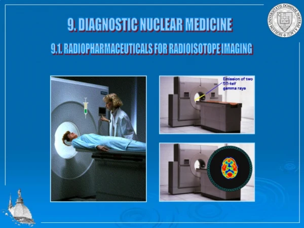
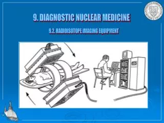
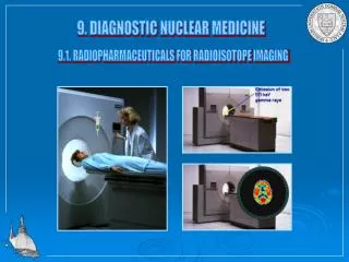
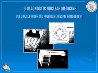
PRINCIPLE OF NUCLEAR MEDICINE. • It uses the principle that a certain radiopharmaceutical (tracer) will at a certain point in time have a preferential uptake by a particular body or tissue. • This uptake is then imaged by the use of detectors mounted in gamma cameras or PET (positron emission tomography) devices.
ADVANTAGES OF NUCLEAR MEDICINE • Ability to image the whole body in a single sitting at a comparably short time hence increased patient through fare. • Minimal radiation burden to patients, personnel and environment- the dosages used are very small • Non-invasive • Able to give information on cellular activity at an early stage.
DISADVANTAGES • Uses very costly and sophisticated equipment. • Requires highly trained personnel. • Higher chances of radiation exposure since the source is unsealed. • Radiation accidents are more likely to occur if safety precautions are compromised.
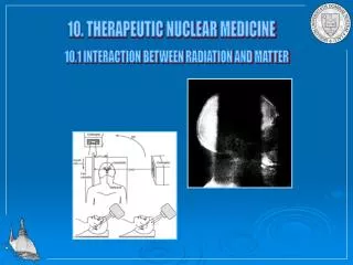
WHAT IS PECULIAR ABOUT NUCLEAR MEDICINE? • Unlike other radiation applications for medical use, nuclear medicine uses unsealed sources of radiation. The tracer is introduced into the body of the patient through several routes (oral, intravenous, percutaneous, intradermally or inhalation) and he/she becomes the source of radiation. • This radiation can then be detected (for diagnostic use) or used to kill some selected unwanted body cells (therapeutic use).
TYPES OF DIAGNOSTIC PROCEDURES DONE AT KNH 1.Whole body radionuclide bone scan -To detect skeletal metastases from cancers such as breast, lung, colon, thyroid, prostate. -To detect the skeleton as the primary source of cancer. -to detect inflammatory conditions of the connective tissue including infections.
cont… -to detect degenerative joint diseases such as osteoarthritis. -to detect sport injuries such as stress fractures -to determine whether pain from a joint prosthesis is due to loosening or infection. -to detect areas of ectopic calcification
Cont… 2.Radionuclide thyroid scan - To determine the anatomical position of the thyroid gland -To determine the functional status of the thyroid gland i.e hypo or hyper thyroidism -To determine possibility of cancer of the thyroid gland (“cold’’ areas) - To determine outcome of radioiodine therapy
Cont… 3.Renal scans -Renogram :will delineate the function of each kidney and the associated collecting system to provide the so-called split kidney function -Renal perfusion studies :eg after kidney transplant -Renal function :glomerular filtration rate (GFR) -Renal anatomy :e.g. ectopic kidneys
Cont… 4.Myocardial perfusion imaging :(MPI) -For diagnosis of coronary artery disease (CAD) -To differentiate between viable and non-viable myocardial tissue e.g. after a myocardial infarction (MI) hence deciding on mode of therapy (surgery vs. medical intervention) -surveillance for those patients known to be at risk of developing CAD eg diabetes
Cont… 5. Lung perfusion studies :to detect pulmonary embolism. 6. Adrenocortical imaging :to detect pheochromocytoma 7. Brain perfusion studies :to detect epileptic foci, dementias, infarction, Parkinsonism.
Cont… 8. Sentinel lymph node (SLN) mapping and lymphoscintigraphy :especially in breast cancer and melanoma. Will help in determining whether lymphoedema is primary ( congenital ) or secondary ( obstructive ). 9. Hepatobiliary (HIDA) Scan: to determine the anatomical and functional integrity of the liver and the biliary ducts; this scan is of great help in differentiating between biliary atresia (whose management is surgical) and other forms of neonatal jaundice (medical management).
2611 patients have undergone diagnostic nuclear medicine studies up to date ( 10 th March 2012 ) as follows;
Therapeutic radiopharmaceuticals • Non-specific • Sr-89, Sm-153, Re-189 • Bone pain palliation • Specific • I-131 • Thyroid cancer, as specific diagnostic if tumor significantly accumulates • Y-90 • Zevalin – monoclonal antibody for B-cell lymphomas
CHALLENGES • Delays in procurement of radiopharmaceuticals e.g 99m-Tc generators and radio-iodine from south Africa • Lack of adequate awareness of the existing NM facilities by our colleagues and the public. • Inadequate numbers of trained personnel and equipment • Lack of PET device and PET/CT hybrid devices for image fusion due to the costs involved • Global shortage of Molybdenum -99.
ACKNOWLEDGEMENTS • IAEA & KNH administration for their supportive roles in making nuclear medicine in this country a reality. T H A N K Y O U.