Bone
General characters:<br>STRUCTURES OF BONE<br>Osteogenic (Osteoprogenitor) cells<br>

Bone
E N D
Presentation Transcript
Histology Bone Dr. M. El-Nagar
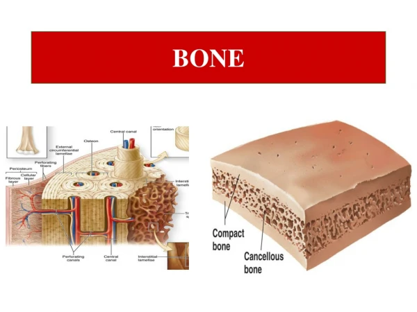
Histology Dr M. El-Nagar Bone • Bone is a specialized dense hard vascular connective tissue. General characters: Like all connective tissues. it consists of cells, fibers and ground substances. Its matrix is hard because it is calcified with mineral salts (mainly calcium and phosphorus) The bone is a highly vascular tissue. It is a dynamic tissue being continuously formed and remodeled under control of varying physiological environmental and hormonal activities. N.B.Similar to cartilage, bone is also a special form of connective tissue. In contrast to cartilage. minerals accumulate and are deposited in the cartilage matrix causing calcification of the developing bones. Consequently. bones become hard and can bear more weight, serve as a rigid skeleton for the body, and provide attachment sites for muscles and organs. STRUCTURES OF BONE Histologically, bone tissue is formed of: Periosteum Endosteum Bone matrix Bone cells Periosteum Periosteum covers the outer surface of bone. It consists of two layers: 1
Histology Dr M. El-Nagar ✓Outer fibrous layer: This layer contains dense collagen fibers, fibroblasts and blood vessels. ✓Inner cellular layer: It is a cellular layer containing fibroblast-like cells called osteogenic cells. When these cells are stimulated, they divide and give rise to osteoblasts. Functions: Provides attachment to tendons, ligaments and muscles. Coarse collagen fiber bundles (Sharpey's fibers) extend from the outer fibrous layer and penetrate the bone tissue where they are continuous with the collagen fibers of the extracellular matrix. Its blood vessels penetrate the matrix to carry oxygen and nutrition. Shares appositional growth and helps repair after fracture. Endosteum Endosteum lines the internal cavities of the bone (bone marrow cavities). It is composed of a single layer of flattened osteogenic cells and osteoblasts. Function: Helps bone growth and repair. Bone Matrix The extracellular substance of bone which is responsible for its rigidity is formed of two major components: Organic component (osteoid): Represents about 35% of the bone formed of: Collagen fibers type I: Form more than 90% of the organic component and is arranged in lamellae. Ground substance: Form 10% of the organic component and includes: ❖Glycosaminoglycan: chondrotin and keratan sulphate. hyaluronic acid is also present. 2
Histology Dr M. El-Nagar ❖Bone matrix proteins: osteocalcin and osteopontin. Inorganic components: Inorganic elements form about 65% of its dry weight. It is composed mainly of calcium and phosphorus. Bicarbonate, citrate, magnesium, potassium and sodium are also present. Bone Cells Bone contains four types of cells. ➢Osteoprogenitor cells ➢Osteoblasts ➢Osteocytes ➢Osteoclasts Osteogenic (Osteoprogenitor) cells Origin: Undifferentiated mesenchymal cells and have the ability to divide. Site: In the deep layer of the periosteum and endosteum. LM: They are small spindle shaped cells with oval nuclei and pale basophilic cytoplasm. EM: Scanty cytoplasm contains free ribosomes, small Golgi apparatus and profiles of RER Functions: In bone growing and fractures, osteogenic cells are stimulated, divide and differentiate to give rise to osteoblasts. 3
Histology Dr M. El-Nagar Osteoblasts Origin: Osteoprogenitor cells. Site: Located at the surface of bone tissues surrounded by matrix, side by side; in a way resembling simple epithelium. LM: They are non-dividing cells. They are large ovoid or polygonal cells. Nucleus: oval and eccentric. Cytoplasm: •Deeply basophilic. •PAS positive granules which are rich in alkaline phosphatase EM: Protein secreting cells: contain extensive rER (which account intense basophilia). mitochondria, well developed Golgi apparatus and numerous secretory vesicles. Function (bone builder cell) ❖Responsible for synthesis of the organic components of bone matrix (collagen type I. Proteoglycan and glycoprotein). ❖Deposition of inorganic components of bone matrix. ❖When osteoblasts are housed in bone lacunae, they become osteocytes. ❖They have receptors for parathyroid hormone by which it secretes osteoclast stimulating factor that activates osteoclasts to resorb bone. 4
Histology Dr M. El-Nagar Osteocytes Origin: Arise from osteoblasts. Site: Present singly in bone lacunae. LM: Appear smaller, branched, and paler than osteoblast. Each cell is imprisoned inside one lacuna (not divide). Cell bodies send many thin processes that pass through corresponding channels or canliculi in the matrix. Nucleus: oval. Cytoplasm: pale basophilic. EM: The cytoplasm is poor in organelles, more condensed peripheral chromatin. Cell processes of neighbouring osteocytes are in contact at their ends to form gap junction to allow flow of ions and possibly molecules from cell to cell. Function: (bone maintenance cell) It is the principal cell of mature bone. Osteocytes are involved in the maintenance of bone matrix. Keep the matrix hard by depositing calcium salts according to body need. 5
Histology Dr M. El-Nagar Osteoclasts Non dividing motile cells. Origin: They arise from monocytes as other phagocytic cells. Site: Motile cells present in shallow cavities known as Howship's lacunae on bone surface at sites of active bone resorption. LM: They are large cells (up to150 μm) Nucleus: multinucleated (up to 50 nuclei). Cytoplasm: acidophilic and strongly positive for acid phosphatase (many lysosomes). The surface of osteoclasts adjacent to the bone shows striations that were interpreted as a brush border. EM: Active osteoclasts (in bone resorption) are subdivided into four morphological zones: Ruffled border zone : ➢The portion of the cell that is directly involved in bone resorption. ➢Its finger-like processes that project into the matrix surface. Clear zone: ➢Peripheral to the ruffled border is a ring-shaped region. ➢It lacks organelles but contains many actin filaments. ➢The plasma membrane of this region is so closely applied to the bone that it forms thesealing zone. 6
Histology Dr M. El-Nagar Vesicular zone: ➢Membrane bounded vesicles of various shapes and sizes are found between the basal and clear zones. ➢They may extend deeply into the cytoplasm between the ruffled border processes. Basal zone: ➢Farthest away from the bone surface is the basal region of the cell it contains its nuclei. numerous mitochondria, and multiple Golgi stalks. Function: (bone eater cell) Osteoclasts are bone eaters. They secrete excess H* ions which produce an acidic medium causing decalcification of the bone. The lysosomal enzymes cause lysis of the organic matrix (collagenases, acid phosphatases and proteolytic enzymes CLASSIFICATIONS OF BONE Classification according to maturity: 1.Primary (immature or woven) bone: The first bone tissue to appear in the fetus. It has irregular or random arrangement of collagen fibers with abundant bone cells. The mineral content is much less than mature form. Later, it is replaced by mature bone except in certain areas e.g. tooth alveoli and insertion of some tendons. It can be also found where there is need for rapid bone formation e.g. following facture. 7
Histology Dr M. El-Nagar 2.Secondary (mature or lamellar) bone: It is the mature and permanent form of bone. The matrix is more abundant and more calcified. It is characterized by concentric regular parallel arrangement of collagen fibers. Osteocytes are arranged in these concentric lamellae at regular intervals. Histological Classification: According to bone lamellar arrangement, secondary bone is classified into two types: ✓Compact Bone: ✓Spongy (Cancellous) Bone. Compact Bone Sites: It is present where support and strength are needed. Shafts of long bones. Inner and outer tables of flat bones e.g. the skull (outer and inner tables of compact bones enclosing a thin layer of spongy bone called diploe). General Organization: it is a mature hard-organized bone tissue. The external surface is covered by periosteum. The bone marrow cavity of the shaft of the long bone is lined by endosteum. The lamellar bone is formed of osteocytes inside lacunae and canaliculi embedded in calcified collagen fibers. Between periosteum and endosteum there are 4 lamellar systems: 1.Outer circumferential lamellae. 8
Histology Dr M. El-Nagar 2.Haversian systems 3.Interstitial lamellae 4.Inner circumferential lamellae. 1.The outer circumferent lamellae Formed by lamellae running parallel to the surface. It lies immediately beneath the periosteum. 2.Haversian systems (osteons) ➢It is a long cylinder parallel to the long axis of bone to form the main bulk the diaphysis. ➢Lamellar bone arranged in concentric cylinders around a central canal called Haversian canal. ➢Each Haversian canal lined by endosteum. ➢Haversian canal contains blood vessels, nerves and the adjacent canals are connected to each other and to periosteum by Volkmann's canals running perpendicular or oblique to Each Haversian canal is surrounded by 4-20 bonelamellae. ➢In each lamella the fibers are parallel to each other and follow a helical course. ➢Between successive lamellae are lacunae, each with one osteocyte, interconnected by canaliculi containing the cells' dendritic processes, for receiving nutrients and oxygen from the vessels in the central canal. ➢The outer boundary of each osteon is a more collagen- rich layer called cement line. 3.Interstitial lamellae Found between Haversian systems. 9
Histology Dr M. El-Nagar 4.Inner circumferential lamellae These are several lamellae present parallel to the endosteum. Spongy Bone Sites: The ends of the long bones (epiphyses) and around its central bone cavity. The Center of irregular and flat bones e.g. the skull, scapula, and pelvic bones General Organization: It appears as branching bone trabeculae and spicules projecting from the inner surface of compact bone, they are covered by endosteum and enclose bone marrow cavities. There are no complete Haversian systems I.e., the osteocytes are located in the branched bone trabeculae with many bone marrow cavities of irregular size inside. HISTOGENESIS OF BONE (OSSIFICATION) General Rules Undifferentiated mesenchymal cells proliferate and differentiate into osteoblasts in highly vascular environment and to chondroblasts in poorly vascular environment. Cartilage cells and models are replaced by bone cells i.e. cartilage does not change to form bone. Bone formation during embryonic development may be of two types: Intramembranous ossification. Endochondral ossification. 10
Histology Dr M. El-Nagar Intramembranous Ossification • It is the development of bone directly from embryonic mesenchyme which destined to be flat bones. Site: Most of flat bones. Steps: The process entails 4 main steps. 1.Development of primary ossification Center around the 8th week of pregnancy. 2.Calcification. 3.Formation of bone trabeculae. 4.Development of periosteum and endosteum. 1. Development of primary ossification Center: ➢Starts in the Center (highly vascularized area) of mesenchymal membrane. ➢The branched undifferentiated mesenchymal cells migrate, condense, and proliferate in the Center. They become osteogenic cells, which differentiate into osteoblasts. 2.Calcification Osteoblasts increase in number and secrete bone matrix including organic component and matrix vesicles rich in alkaline phosphatase that has a great role in deposition of calcium and phosphorous (mineralization). When osteoblasts are trapped inside lacunae in this calcified matrix and become osteocytes. Collagen fibers are randomly oriented and calcification progress rapidly. 11
Histology Dr M. El-Nagar 3.Formation of bone trabeculae: Bone matrix appears as spicules radiating from the Center of this growing area As this process is going on. the vascular connective tissue between these lamellae is transformed into bone marrow. 4.Development of periosteum and endosteum Remnants of mesenchymal membrane on both outer and inner regions of this developing bone surface change into periosteum while those covering trabeculae form endosteum. The spongy bone deep to the periosteum is transformed into compact bone. forming the outer and inner tables enclosing the spongy bone (called diploe in the skull) Endochondral Ossification It is the development of bone in place of a cartilage model. Sites: Long and short bones which requires the presence of a cartilage model of the same shape. Steps: 1.Development of a cartilaginous model. 2.Growth of this cartilage model through the formation of: Primary ossification Center Secondary ossification Center 12
Histology Dr M. El-Nagar 1.Development of a Cartilaginous Model: At the site where bone will be formed, a cartilaginous model of the same shape develops. This cartilage is surrounded by perichondrium. 2.Growth of Cartilage: Primary Ossification Center: At the perichondrium The perichondrium in the middle of the diaphysis becomes vascularized, so the chondrogenic cells differentiate into osteogenic cells and the perichondrium changes into periosteum. Osteogenic cells differentiate into osteoblast. The newly formed osteoblasts secrete bone matrix and form sub-periosteal bone collar on the surface of the cartilage model. The formed periosteum is invaded by periosteal bud, which is formed of blood vessels, osteoprogenitor cells, osteoclasts and hemopoietic cells. Inside the middle of the cartilage and at the same time the following occur: The chondrocytes at the Center of the model hypertrophy accumulate glycogen and appear vacuolated. Bone collar prevents the diffusion of nutrients to the hypertrophied chondrocytes causing them to die leaving empty lacunae forming large cavities. The periosteal bud enters the cavities in the Center of cartilage model through holes formed at the periosteum by the action of osteoclasts. The osteoblasts and osteoclasts arrange themselves around areas of calcified cartilage matrix. The osteoclasts erode the cartilage matrix while osteoblasts deposit new bone until all cartilage is replaced by bone. 13
Histology Dr M. El-Nagar This bone deposition results in spongy bone formation in the Center of cartilage model which is called primary ossification Center. Osteoclasts resorb the irregular bone trabeculae in the Center to form one central regular bone marrow cavity filled with hemopoietic cells. This Center expands at both ends towards the epiphyses. So, at birth the long bone has bony shaft and two cartilaginous ends. Secondary Ossification Center: It starts at both epiphyses of the developing bone. Similar steps to those occurred in the primary Center progress, however, no bone collar is formed, and bone expands radially. It continues to grow and bone tissue tills the epiphysis except the articular surface (articular cartilage) and the epiphyseal plate. The articular cartilage remains throughout life whereas the epiphyseal plate continues growth till being closed at adulthood and bone length becomes stable. Bone Growth in Length (Epiphyseal Plate): • Five zones can be distinguished under the microscope. Zone of resting (reserve) cartilage Chondrocytes are mitotically active. They are randomly distributed throughout the matrix. Zone of proliferation: Chondrocytes divide and the daughter cells are arranged in rows and columns parallel to the direction of bone growth. 14
Histology Dr M. El-Nagar Zone of maturation and hypertrophy: Chondrocytes mature, hypertrophy, and accumulate glycogen and alkaline phosphatase in their cytoplasm. The matrix between the cells becomes thin. Zone of calcification: Deposition of calcium in the matrix prevents diffusion of nutrients to the hypertrophied chondrocytes. Chondrocytes die leaving empty spaces separated by calcified matrix. Zone of ossification: The osteogenic cells invade this area and differentiate into osteoblasts, which secrete organic matrix and matrix vesicles with subsequent calcification. This results in formation of cancellous bone separated by bone marrow cavities. Osteoclasts erode the irregular bone trabeculae to a regular bone marrow cavity. 15