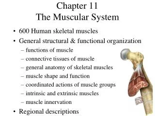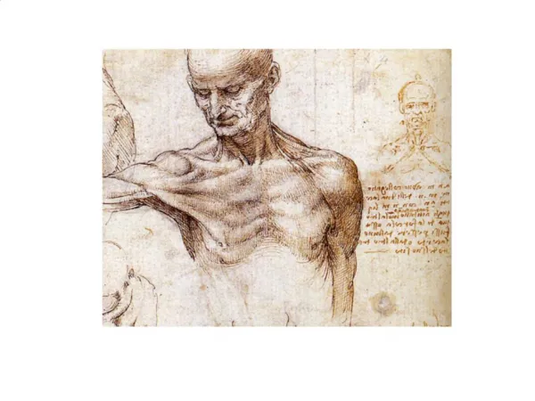
Skeletal Muscles: Function, Naming, and Mechanics
940 likes | 969 Vues
Dive into the intricacies of skeletal muscles, from their functional groups and naming conventions to the arrangement of fascicles. Explore prime movers, antagonists, synergists, fixators, and muscle mechanics. Learn how to name muscles based on location, shape, size, and more. Discover the major skeletal muscles of the body categorized by function and location, along with their origins, insertions, actions, and innervation. Enhance your knowledge of facial, head, shoulder, arm, thorax, abdomen, pelvis, thigh, leg, and neck muscles.

Skeletal Muscles: Function, Naming, and Mechanics
E N D
Presentation Transcript
Skeletal Muscles: Functional Groups • Prime movers • Provide the major force for producing a specific movement • Antagonists • Oppose or reverse a particular movement • Synergists • Add force to a movement • Reduce undesirable or unnecessary movement • Fixators • Synergists that immobilize a bone or muscle’s origin
Naming Skeletal Muscles • Location—bone or body region associated with the muscle • Shape—e.g., deltoid muscle (deltoid = triangle) • Relative size—e.g., maximus (largest), minimus (smallest), longus (long) • Direction of fibers or fascicles—e.g., rectus (fibers run straight), transversus, and oblique (fibers run at angles to an imaginary defined axis)
Naming Skeletal Muscles • Number of origins—e.g., biceps (2 origins) and triceps (3 origins) • Location of attachments—named according to point of origin or insertion • Action—e.g., flexor or extensor, muscles that flex or extend, respectively
Muscle Mechanics: Arrangement of Fascicles • Circular • Fascicles arranged in concentric rings (e.g., orbicularis oris) • Convergent • Fascicles converge toward a single tendon insertion (e.g., pectoralis major) • Parallel • Fascicles parallel to the long axis of a straplike muscle (e.g., sartorius) • Fusiform • Spindle-shaped muscles with parallel fibers (e.g., biceps brachii) • Pennate • Short fascicles attach obliquely to a central tendon running the length of the muscle (e.g., rectus femoris)
(a) (g) (b) (f) (b) Convergent (pectoralis major) Circular (orbicularis oris) (c) (e) (c) Parallel (sartorius) (d) Unipennate (extensor digitorum longus) (d) (e) Bipennate (rectus femoris) (f) Fusiform (biceps brachii) (g) Multipennate (deltoid) Figure 10.1
Major Skeletal Muscles of the Body • Grouped by function and location • Information for each muscle • Name and description—note information in the name • Origin and insertion—there is usually a joint between the origin and the insertion • Action—insertion moves toward origin; best learned by acting out muscle movement on one’s own body • Innervation—name of major nerve that supplies the muscle
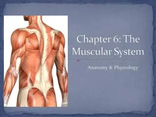
Facial Head Epicranius, frontal belly Temporalis Orbicularis oculi Masseter Zygomaticus Shoulder Orbicularis oris Trapezius Neck Deltoid Sternohyoid Arm Platysma Triceps brachii Sternocleidomastoid Biceps brachii Thorax Brachialis Pectoralis minor Forearm Serratus anterior Pronator teres Pectoralis major Brachioradialis Intercostals Flexor carpi radialis Abdomen Palmaris longus Rectus abdominis Pelvis/thigh Internal oblique Iliopsoas Transversus abdominis Pectineus External oblique Thigh Thigh Rectus femoris Tensor fasciae latae Vastus lateralis Sartorius Vastus medialis Adductor longus Leg Gracilis Fibularis longus Leg Extensor digitorum longus Gastrocnemius Tibialis anterior Soleus Figure 10.4
Neck Epicranius, occipital belly Sternocleidomastoid Arm Trapezius Triceps brachii Shoulder Brachialis Deltoid Forearm Infraspinatus Brachioradialis Teres major Extensor carpi radialis longus Rhomboid major Latissimus dorsi Flexor carpi ulnaris Hip Extensor carpi ulnaris Gluteus medius Gluteus maximus Extensor digitorum Iliotibial tract Thigh Adductor magnus Hamstrings: Leg Biceps femoris Gastrocnemius Semitendinosus Semimembranosus Soleus Fibularis longus Calcaneal (Achilles) tendon Figure 10.5
Muscles of the Head • Two groups • Muscles of facial expression • Muscles of mastication and tongue movement
Muscles of Facial Expression • Insert into the skin • Important in nonverbal communication • All innervated by cranial nerve VII (facial nerve)
Muscles of Facial Expression • Epicranius (occipitofrontalis) • Bipartite muscle consisting of the • Frontalis • Occipitalis • Galea aponeurotica—cranial aponeurosis connecting above muscles • The two muscles have alternate actions of pulling the scalp forward and backward
Epicranius Galea aponeurotica Corrugator supercilii Frontal belly Orbicularis oculi Occipital belly Levator labii superioris Zygomaticus minor and major Temporalis Buccinator Masseter Risorius Sternocleidomastoid Orbicularis oris Trapezius Mentalis Splenius capitis Depressor labii inferioris Depressor anguli oris Platysma Figure 10.6
Muscles of Mastication and Tongue Movement • Four pairs involved in mastication • Prime movers of jaw closure • Temporalis and masseter • Grinding movements • Medial and lateral pterygoids
Muscles of Mastication and Tongue Movement • All are innervated by cranial nerve V (trigeminal nerve) • Buccinator muscles (of facial expression group) also help by holding food between the teeth • Three muscles anchor and move the tongue • All are innervated by cranial nerve XII (hypoglossal nerve)
Temporalis Orbicularis oris Masseter Buccinator (a) Figure 10.7a
Lateral pterygoid Medial pterygoid Masseter pulled away (b) Figure 10.7b
Tongue Styloid process Styloglossus Genioglossus Hyoglossus Stylohyoid Mandibular symphysis Hyoid bone Geniohyoid Thyrohyoid Thyroid cartilage (c) Figure 10.7c
Muscles of the Anterior Neck and Throat • Most are involved in swallowing • Two groups • Suprahyoid • Infrahyoid
Suprahyoid Muscles of the Anterior Neck and Throat • Four deep muscles are involved in swallowing (they move the hyoid bone and larynx) • Form the floor of the oral cavity • Anchor the tongue • Move the hyoid bone and the larynx
Median raphe Anterior belly Mylohyoid Digastric Stylohyoid Posterior belly Hyoid bone Omohyoid (superior belly) Stylohyoid (cut) Thyrohyoid Sternohyoid Thyroid cartilage of the larynx Sternocleido- mastoid Thyroid gland Omohyoid (inferior belly) Sternothyroid (a) Figure 10.8a
Tensor veli palatini Levator veli palatini Styloid process Buccinator Superior pharyngeal constrictor Mandible Middle pharyngeal constrictor Mylohyoid (cut) Hyoid bone Geniohyoid Thyrohyoid membrane Hyoglossus Inferior pharyngeal constrictor Thyroid cartilage of larynx (c) Esophagus Trachea Figure 10.8c
Muscles of the Neck and Vertebral Column • Two functional groups • Muscles that move the head • Muscles that extend the trunk and maintain posture
Muscles of the Neck and Vertebral Column: Head Movement • Sternocleidomastoid—major head flexor • Suprahyoid and infrahyoid—synergists to head flexion • Sternocleidomastoid and scalenes—lateral head movements • Semispinalis capitis—synergist with sternocleidomastoid • Splenius (capitis and cervicis portions): head extension, rotation, and lateral bending
Base of occipital bone 1st cervical vertebra Mastoid process Middle scalene Sternocleido- mastoid Anterior scalene Posterior scalene (a) Anterior Figure 10.9a
Mastoid process Splenius capitis Spinous processes of the vertebrae Splenius cervicis (b) Posterior Figure 10.9b
Muscles of the Neck and Vertebral Column: Trunk Extension • Deep (intrinsic) back muscles • Erector spinae (sacrospinalis) group—prime movers of back extension and lateral bending • Iliocostalis • Longissimus • Spinalis • Semispinalis and quadratus lumborum—synergists in extension and rotation
Ligamentum nuchae Mastoid process of temporal bone Semispinalis capitis Longissimus capitis Iliocostalis cervicis Semispinalis cervicis Longissimus cervicis Semispinalis thoracis Iliocostalis thoracis Longissimus thoracis Spinalis thoracis Iliocostalis Erector spinae Longissimus Spinalis Multifidus Iliocostalis lumborum Quadratus lumborum External oblique (d) Figure 10.9d
Muscles of the Thorax • Muscles of respiration • External intercostals—more superficial muscles that elevate ribs for inspiration • Internal intercostals—deeper muscles that aid forced expiration • Diaphragm • Partition between thoracic and abdominal cavities • Most important muscle in inspiration • Innervated by phrenic nerves
External intercostal (a) Internal intercostal Figure 10.10a
Xiphoid process of sternum Foramen for inferior vena cava Foramen for esophagus Costal cartilage Central tendon of diaphragm Diaphragm Foramen for aorta Lumbar vertebra 12th rib Quadratus lumborum Psoas major (b) Figure 10.10b
Muscles of the Abdominal Wall • Four paired muscles; their fasciae and aponeuroses form the lateral and anterior abdominal wall • Internal obliques • External obliques • Transversus abdominis • Rectus abdominis
Pectoralis major Serratus anterior Linea alba Tendinous intersection Transversus abdominis Internal oblique Rectus abdominis External oblique Inguinal ligament (formed by free inferior border of the external oblique aponeurosis) Aponeurosis of the external oblique (a) Figure 10.11a
Muscles of the Abdominal Wall • Fascicles of these muscles run at angles to one another, providing added strength • All are innervated by intercostal nerves • Actions of these muscles • Lateral flexion and rotation of the trunk • Help promote urination, defecation, childbirth, vomiting, coughing, and screaming
Rectus abdominis Internal oblique External oblique Lumbar fascia IIiac crest Pubic tubercle Lumbar fascia Transversus abdominis Inguinal ligament (b) Figure 10.11b
Muscles of the Pelvic Floor • Pelvic floor (pelvic diaphragm) is composed of two paired muscles • Levator ani • Coccygeus • Both are innervated by sacral nerves
Muscles of the Pelvic Floor • Functions of the pelvic diaphragm • Seals the inferior outlet of the pelvis • Supports pelvic organs • Lifts pelvic floor to help release feces • Resists increased intra-abdominal pressure
Muscles of the Perineum • Urogenital diaphragm • Anterior half of perineum, inferior to pelvic floor • Deep transverse perineal muscle • External urethral sphincter (voluntary control of urination)
Anterior Symphysis pubis Pubococcygeus Levator ani IIiococcygeus Urogenital diaphragm Urethra Vagina Anal canal Obturator internus Coccyx Levator ani Piriformis Coccygeus Pelvic diaphragm Posterior (a) Figure 10.12a
Urethral opening External urethral sphincter Vaginal opening Inferior pubic ramus Deep transverse perineal muscle Central tendon Anus External anal sphincter Male Female (b) Figure 10.12b
Superficial Muscles of the Thorax • Most are extrinsic shoulder muscles • Act in combination to fix the shoulder girdle (mostly the scapula) and move it to increase range of arm movements • Actions include elevation, depression, rotation, lateral and medial movements, protraction, and retraction
Superficial Muscles of the Thorax • Anterior extrinsic shoulder muscles • Pectoralis minor • Serratus anterior • Subclavius • (Pectoralis major considered later with muscles that act on the humerus)
Subclavius Sternocleido- mastoid Clavicle Subscapularis Deltoid Pectoralis minor Pectoralis major Coracobrachialis Sternum Serratus anterior Biceps brachii Humerus (a) Figure 10.13a
Superficial Muscles of the Posterior Thorax • Posterior extrinsic shoulder muscles • Trapezius • Levator scpulae • Rhomboids (major and minor) • (Latissimus dorsi considered later with muscles that act on the humerus)
Levator scapulae Trapezius Supraspinatus Clavicle Deltoid Spine of scapula Rhomboid minor Infraspinatus Teres minor Rhomboid major Teres major Humerus Latissimus dorsi (c) Figure 10.13c
Muscles Crossing the Shoulder Joint • Nine muscles cross the shoulder joint to insert on and move the humerus • Three are prime movers of the arm • Pectoralis major • Latissimus dorsi • Deltoid • Actions include flexion, extension, adduction, abduction, and rotation of humerus
Muscles Crossing the Shoulder Joint • Four muscles are rotator cuff muscles • Supraspinatus • Infraspinatus • Teres minor • Subscapularis • Reinforce the capsule of the shoulder • Act as synergists and fixators • Two additional muscles are synergists: coracobrachialis and teres major
Clavicle Deltoid Sternum Pectoralis major Coracobrachialis Triceps brachii: Lateral head Long head Medial head Biceps brachii Brachialis Brachioradialis (a) Anterior view Figure 10.14a
Supraspinatus* Spine of scapula Deltoid (cut) Greater tubercle of humerus Infraspinatus* Teres minor* Teres major Triceps brachii: Lateral head Long head Latissimus dorsi Humerus Olecranon process of ulna Anconeus (b) Posterior view * Rotator cuff muscles Figure 10.14b