Understanding the Interrelationship Between Form and Function in Animal Biology
420 likes | 553 Vues
This overview explores the critical relationship between form and function in biological structures, particularly in animals. It highlights that the configuration of a structure often determines its capabilities, reflecting evolutionary adaptations. The discussion covers four primary tissue types: epithelial, connective, muscle, and nervous tissue, each playing vital roles in organism functionality. Additionally, we examine the integumentary system and skeletal system, detailing their respective forms, functions, and importance for overall biological health and activity.

Understanding the Interrelationship Between Form and Function in Animal Biology
E N D
Presentation Transcript
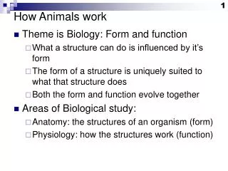
How Animals work • Theme is Biology: Form and function • What a structure can do is influenced by it’s form • The form of a structure is uniquely suited to what that structure does • Both the form and function evolve together • Areas of Biological study: • Anatomy: the structures of an organism (form) • Physiology: how the structures work (function)
We are a tube within a tube • We are a thick walled tube • Running through the center of the tube is a digestive system, another tube. http://biology.kenyon.edu/courses/biol112/Biol112WebPage/Syllabus/Topics/Week%207/Resources/coelom.GIF http://www.mscd.edu/~biology/231course/cavity.jpg
Which way is which? http://anthro.palomar.edu/animal/images/dorsal.gif
Tissues • A tissue is a group of similar cells that usually have a similar embryological origin and are specialized for a particular function. • Tissue cells are often separated by non-living, intercellular materials that cells produce. This substance is called the matrix. • Histology – the study of tissues
Four Principal Types of Tissues: • Epithelial –covering and lining; and glands • Connective – protects and supports, binds organs together, stores energy, and provides immunity • Muscle – movement • Nervous –transmits impulses that coordinate body activities
Feature of Epithelial Tissue • Closely packed cells with little extracellular material • Epithelial cells are arranged in continuous sheets, in single or multiple layers • Epithelial cells have two surfaces • One side is up against other tissues • The other side faces an open space http://www.cnas.missouristate.edu/labimages/biology/bio122/images/Week%208%20Images/trachea.jpg
Connective Tissue • Connective tissue is the most abundant and widely distributed tissue in the body . • The functions of connective tissue: • Binding, attachment and support (bones, tendons and ligaments, organs) • Protection- bones and adipose tissue • Insulation - adipose tissue • Storage - of energy and fat soluble substances • Transportation - blood
Muscle Tissue Skeletal Smooth Cardiac voluntary involuntary involuntary cells are long short, spindle-shaped branched many nuclei single nucleus single nucleus striated nonstriated striated intercalated disks Diagram of skeletal muscle
Nervous tissue Sends signals to other parts of body neurons : Dendrites - (one or many) receive incoming signals Cell body Axon - (one) transmits outgoing signals neuroglia : protective and supporting cells
Axon dendrites
Organs are two or more tissues which together perform a specialized function. • Epithelial membranes are thin structures that usually contain both epithelial and connective tissue.
Integumentary system is the skin and the organs derived from it (hair, glands, nails) • One of the largest organs • 2 square meters; 10-11 lbs. • Largest sense organ in the body • The study of the skin is Dermatology
Three types of epithelial membranes • Serous Membranes • Line cavities and cover organs • Mucous membranes • Line cavities that open to the exterior • Layer of epithelium over connective tissue; • Cutaneous membrane is the skin • the major organ of the integumentary system
Functions: 1. Regulation of body temperature • Cellular metabolism produces heat as a waste product . • High temperature • Dilate surface blood vessels • Sweating • Low temperature • Surface vessels constrict • shivering
2. Protection physical abrasion dehydration ultraviolet radiation 3. Sensation touch vibration pain temperature
4. Excretion • Sweat, rid excess salt for example • 5. Immunity/ Resistance • Keeps microbes out, fights infections • 6. Blood Reservoir • 8-10 % of blood in a resting adult • 7. Synthesis of vitamin D • Produced by exposure to UV light • aids absorption of calcium
Anatomy • Epidermis Skin • Dermis • Subcutaneous layer or hypodermis See next two slides
The Skeletal System • comprised mostly of connective tissue • In the case of most vertebrates, a skeletal system is comprised of bone • Various bone types
Functions of Bone • Support • For soft tissues • For attachment of skeletal muscles • Protection • Movement • Storage : minerals, fat • Blood cell production – hematopoiesis • Erythrocytes, leukocytes, platelets
Matrix • As a connective tissue, bone is living cells embedded in a matrix consisting of • Secreted substances (proteins) • Proteoglycans and glycoproteins • Collagen – reinforces, gives tensile strength • Mineral salts • Tricalcium phosphate (hydroxyapatite) and calcium carbonate • Calcium is thus needed for bone. • Bone is an active, living material!
Remodeling • Bone is continually being broken down and reformed • 3-5 % of bone calcium exchanged each year • In response to stress on bones • Balance between actions of osteoclasts and osteoblasts • Osteo means bone • Osteoblasts MAKE bone, osteoclasts dismantle it. • Vitamins needed for maintenance, esp. D
3 related organ systems • Two important things that animals do: feed and breed • We’ll skip “breed”, but explore 3 organ systems by looking at “feed” • Part of “survival of the fittest” is being able to procure nutrients to survive and live long enough to breed. • Digestive system: processes nutrients • Respiratory exchanges nutrients and wastes • Cardiovascular system transports these
Needed to function (but not discussed) • Urinary system: without kidneys to filter waste products from the blood for excretion, we’d be in trouble. • Muscles: movement required for animals. • Nervous system and endocrine system: • nerve cells allow communication by electrochemical signals • Coordinated with chemical signal: hormones
Digestive system • Food is needed • Organic materials to break down for energy • Organic molecules as building blocks for cellular material • Specific organic molecules (amino acids, vitamins) we are unable to make ourselves • Certain minerals needed to assist enzymes • How much? • We need enough energy (measured in calories) to power our activities. • Over and under nutrition can occur.
A digestive system needed • A large organism has a system for extracting nutrients from food for distributing to the rest of the body. • Food must be physically broken down • Food must be chemically broken down • Nutrients must be absorbed • Excess water used in digestion must be recovered • Waste must be excreted
Parts of a digestive system • Mouth • Teeth for breaking up food • Saliva for wetting, breakdown of starch • Esophagus: muscular tube for transferring food to the stomach • Stomach • Physical digestion; muscular stomach churns • Chemical: low pH (high stomach acid) dissolves food; enzymes also breakdown food
Food passes into small intestine • Bile salts and enzymes from liver (via gallbladder) act as detergents, break up fats. • Pancreas neutralizes stomach acid, adds enzymes that break down food more. • Small intestine: site of nutrient absorption • Possesses a high surface area • Cells are arranged in villi • Each cell is covered with microvilli • Each villus contains lots of blood capillaries for transporting nutrients away
Form and function in the small intestine villi High surface area for lots of absorption. http://users.rcn.com/jkimball.ma.ultranet/BiologyPages/V/villi.gifhttp://distance.stcc.edu/AandP/AP/imagesAP2/digestion/microvilli.jpg
The end of the line • Large intestine = colon • Most of the water is absorbed here • In an irritated colon, material is moved through quickly before all the water is absorbed: diarrhea • Home to billions of bacteria • Most are anaerobic • Responsible for bad odors, production of gas from fermentation • Feces are mostly bacterial bodies; undigested food a smaller proportion
Respiratory system • During metabolism, cells “burn” organic molecules. • E.g. glucose ultimately gets converted to CO2, energy is released. • Process needs oxygen, releases CO2 • Respiratory system must efficiently get O2 into the body • Yet protect from microbes, dirt and dust, drying out, etc.
Respiratory anatomy • Nose, sinuses • Trachea = windpipe, tube that leads from oral cavity (mouth) to lungs • Bronchi: the trachea branches • Bronchioles: the bronchi branch into smaller tubes that lead to the lobes of the lung • Alveoli = air sacs: small compartments in which gasses are transferred between air and bloodstream
Lungs http://www.aduk.org.uk/gfx/lungs.jpg
Respiratory protections • Ciliated epithelium • Surfaces lined with cells that secrete mucus, push mucus along with cilia • Mucus with entrapped bacteria, dust, pushed towards mouth for coughing out or swallowing • Swallowing leads to stomach, acid • Things that kill epithelium interfere with these protections • Influenza virus • smoking
Single ciliated cell Cilia http://n2.bioeng5.bioeng.auckland.ac.nz/ontology/images/RespiratoryEpitheliumCiliatedEpithelialCell.png
Where does Oxygen go? http://training.seer.cancer.gov/module_anatomy/images/quiz_dd_pulmonary_circulation.jpg
Cardiovascular terms to know • Cardio means heart; vascular means tubes • From big to small • Arteries, arterioles, capillaries: • Take blood AWAY from the heart • Veins, venules, capillaries • Bring blood TO the heart • Capillaries are where exchange of materials between blood stream and cells occurs.
Status of blood • Pulmonary circulation • Blood traveling AWAY from heart to lungs has little O2 content (in blue): pulmonary artery • Blood traveling from lungs TO heart has lots of O2 (in red): pulmonary vein.
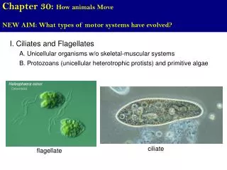
The rest of the circulation • Heart has 4 chambers in a mammal • 2 atria and 2 ventricles • Pulmonary and systemic circulation • Blood returns from body into right atrium • Flows into right ventricle, pumped to lungs • Returns from lungs to left atrium • Flows into left ventricle, pumped to the rest of the body • Main artery leading to rest of body: aorta
Heart structure Ao = aortaLA = left atriumLV = left ventriclePA = pulmonary arteryRA = right atriumRV = right ventricle Match up with description on previous slide. http://www.rch.org.au/cardiology/media2/Fontan_pic1.gif