Download
1 / 68
680 likes | 838 Vues
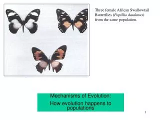
Genetics: The source of variability for evolution. How population survival strategies determine human biology and provides the basic background for human variation. Diversity of form and function. The basis of evolution is variation

E N D
Genetics: The source of variability for evolution How population survival strategies determine human biology and provides the basic background for human variation
Diversity of form and function • The basis of evolution is variation • But, where does variability in biological form and function come from? • There are two levels of evolution we will be interested in: • Macro-level evolutionary change, the appearance of new species, and • Micro-level evolutionary change, the generation by generation changes in the genes of populations.
What does the genetic material do, anyway? • The genetic material has a number of important functions: 1. Transmit genetic information from one generation to the next (humans produce human infants and not rats or elephants). 2. Since every cell in the body (with several exceptions) has more or less the same genetic material as the original cell (the fertilized egg), the genetic material must be able to reproduce itself when new cells are produced during growth and development as well as normal body maintenance. 3. The genetic materials are organized around a sequence of chemical ‘bases’ that encode for the synthesis of proteins, a huge class of chemicals that perform a wide range of functions in the body.
What determines cell structure and function? • Proteins that are expressed • Unique expression by cell type • How is this controlled? • Look to the cell nucleus
Chromosomes • Carries information as part of their structure • Name=colored bodies when stained and seen microscopically • Species-specific number in each cell nucleus, with the chromosome number usually expressed in pairs (the complexity of the living thing is not reflected in the chromosome number (chimps, for example, have more chromosomes than humans).
Human chromosomes • Species specific number=46 • 23 pairs of chromosomes • Specifially: • 22 pairs of autosomes • or, homologous chromosomes • 1 pair of sex chromosomes • XX female • XY male • Question: Why are there pairs of chromosomes?
Where do the chromosomes come from? • We are originally one cell: • 23 of maternal origin • ova carry these • 23 of paternal origin • sperm carry these • If every cell has 46, how do these end up with only 23 and why?
Meiosis • How many of you remember the process of meiosis well enough to explain it to your classmates?
If we start out as one cell, how do we get so big and complicated? • BIG =cell division (Mitosis) • Complicated =cell differentiation
Mitosis • Cell Division • Chromosomal Replication
Differentiation • Unique proteins in different cell types
Proteins: What are they? • You are what you eat! • Functions include: • Structure • Transport • Immune • Function reflects their structure • Proteins have 3 dimensional structure • Folded chains
Proteins: Structural specifics • Structure: • Three dimensional • Folded chain • Polypeptide chain • of amino acids (aa) • 20 common aa • Different proteins have different aa sequences
Chemical group based on their composition: an “amine” and an “acid” Of the 20 common aa, 10 the body can make 10 must be eaten (essential aa) Glycine (gly) Glutamic acid (glu) Alanine (ala) Aspartic acid (asp) Valine (val) Isoleucine (Ile) Leucine (leu) Serine (ser) Threonine (thr) Proline (pro) Lysine (lys) Arginine (arg) Glutamine (gln) Aspargine (asn) Methionine (met) Cysteine (cys) Tryptophan(trp) Tyrosine (tyr) Histidine (his) Phenylalanine(phe) Amino acids: What are they and where do they come from?
Proteins: How they are made: 1. From amino acids • Polypeptide chains=aa • Sequence of aa crucial to structure, and thus function • Sequence determined by series of nucleic acids and the genetic code • Determined by a gene met val his leu thr asp ala glu lys val ala ala ss cys leu trp gly lys val asn ser asp glu
What is a gene? • A “recipe” for a protein • Located at a specific region (locus) on a specific chromosome • Implications: • different chromosomes carry different information • Question: • do homologous chromosomes carry the same information?
A gene up-close: is a coding sequence of DNA • The relationship between chromosomes and DNA: • Chromosomes are packaged DNA
DNA • Double helix structure • Biochemically: • Deoxyribose sugar • NucleicAcids • purines: adenine, guanine • pyrimidines: thymine, cytosine • Base pair rules: c g a t
For example, how is a piece of coding DNA translated? CCTGAGGAG GGACTCCTC Genes and their protein products:How does a gene “code” for a protein?What is the process by which the structure of DNA determines the structure of a protein?
The genetic code • 1. Only one strand of DNA is the ‘recipe’, or code • The “genetic code” : three sequential nucleic acids specify an amino acid • DNA: CAAGTAGAATGCGGACTTCTT • AA: val his leu thr pro glu glu
Code to Protein: Shuttle system • A messenger transmits DNA sequence to protein assembly site • messenger RNA (Ribose Nucleic Acid) • distinct from DNA: single strand C G A Uracil • self-assembles as it “reads” the DNA by base-pair rules • goes to ribosome, site of protein assembly
Translate this DNA into mRNA: • CAAGTAGAATGCGGACTTCTT • A. GTTCATCTTACGCCTGAAGAA • B. GUUCAUCUUACGCCUGAAGAA
The genetic code: codons • mRNA: GUUCAUCUUACGCCUGAAGAA • GUU CAU CUU ACG CCU GAA GAA
Mirror image of DNA Identifies a sequence of amino acids, and thus a protein HOW Are amino acids gathered together in the correct sequence? The genetic code: A TRANSLATOR molecule t-RNA: M-RNA strand to protein Amino Acid 3NA
m-RNA meets t-RNA:polypeptide chain of amino acids built • mRNA:GUUCAUCUUACGCCUGAAGAAAAG • GUU CAU CUU ACG CCU GAA GAA AAG caa gua gaa ugc gga cuu cuu uuc leu thr pro lys val his glu glu Beta Globin (146 aa)
Case Study: Genetics in action at the level of the population • Case study: SCA • Background: • 1912 James Herrick: • Case Report Blood smear analysis • 1940’s family studies: • Mendelian genetics
Proteins: Structural specifics • Structure: • Three dimensional • Folded chain • Polypeptide chain • of amino acids (aa) • 20 common aa • Different proteins have different aa sequences
Red Blood Cells: What do they do? • Origin in bone marrow • 120 day life cycle • Oxygen-carriers • Pick up oxygen in lungs • Deliver oxygen to body tissues • By what mechanism? Rbcsinblood on top half alvertonoutpouch on bottom
A Protein!Hemoglobin: Function • In red blood cells • Transport protein • Carries oxygen • How?
Three dimensional Four components: Two “alpha” chains chromosome 16 Two “beta” chains chromosome 11 Red marks the spot! Where oxygen binds Iron ion critical here Hemoglobin: Structure The function depends on structure: How hemoglobin works
Sickle Cell Anemia • Sickle Cell: • red blood cell shape • Anemia: • poor oxygen delivery • Cause: • abnormal hemoglobin • A genetic disease
What causes the sickling? • Hemoglobin molecule changes shape • Results in distortion of rbc • Functional effects?
Why does the hemoglobin do this? • WHEN: Abnormal hemoglobin molecule unstable under conditions of low oxygen, high acidity • HOW: Crystalline • structure results • WHY? Structural instability
First 6 amino acids: Beta globin gene: 146 amino acids Hbs beta globin chain one different amino acid valine replaces glutamic acid at position 6 Hemoglobin “S” vs Hemoglobin “A”(Sickle [S] vs Normal [A]) Valine Valine Histidine Histidine Leucine Leucine Threonine Threonine Proline Proline Glutamic acid Valine A S
One nucleic acid apart • DNA • HbA: GGACTTCTT • HbS: GGACATCTT Genetic Pro Glu Glu Mutation Pro Val Glu
Population Frequency of HbS Heterozygote vs Homozygote? • Africa • In some places, 1 in 5 people are carriers, or • HbS/HbA genotype, or heterozygous (hetero=different) • A co-dominant trait: both proteins are expressed Dominant vs recessive?
Review: • At each locus, there are two genes; they are either the same or different. This illustrates allelic variability • Homozygote • Heterozygote • Dominant • Recessive
HbS and adaptation: • In a population of 100 individuals, calculate the number of HbS and HbA genes if 20 % of the people are heterozygotic and the rest are homozygotic normal. • What is the percentage of HbS and HbA genes in the population? • Why do you think there are no HbS/HbS individuals?