Download
1 / 42
471 likes | 1.53k Vues
Surgical pathology of the appendix. Acute appendicitis Chronic appendicitis Tumors of the appendix. Appendix. Functions – not clear in humans - it may have a significance in immune defense – abundance of lymphoid follicles

E N D
Surgical pathology of the appendix Acute appendicitis Chronic appendicitis Tumors of the appendix
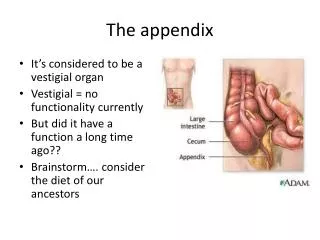
Appendix • Functions– not clear in humans- it may have a significance in immune defense – abundance of lymphoid follicles - removal of the appendix may be a cause for an increase in colonic cancer incidence - not supported by controlled studies - endocrine function
Typical position • 2.5 cm bellow the ileo-cecal valve (base of appendix) the only fix region – important when trying to find the appendix • Taeniae converge at the base of the appendix • 84% free mobile in any possible location • 16% fixed retrocecal
Acute apendicitis • Essentials of diagnosis • Abdominal pain • Anorexia, nausea, vomiting • Localized abdominal tenderness • Low grade fever • Leukocytosis
General considerations • = acute inflammation of the appendix wall that starts in the mucosa and may extend to adjacent organs • 70% of cases present obstruction of the proximal lumen: • Fibrous bands, fecaliths, foreign bodies • Tumors, parasites, lymphoid hyperplasia • External compression • Inflammation starts in the mucosa with ulcerations and secondary bacterial infection
Close tube • Blood supply affected as disease progresses • Infection in the wall • Increased pressure • Puss formation inside the lumen • Wall destruction: gangrene + perforation • Bacterial peritonitis may be limited by adhesions (plastic peritonitis)
Clinical findings • Protean manifestation: may mimic a variety of conditions • Progression of symptoms is essential
Clinical findings • Onset: vague abdominal discomfort • Followed: • Nausea, anorexia, indigestion • Vomiting • Pain, mild, localized in the epigastrum • Pain: localized in RLQ + • Pain or discomfort (moving, walking, coughing)
Examination • At this moment: • Tenderness on coughing, localized in RLQ • Localized tenderness on palpation • Slight muscular rigidity • Rebound tenderness referred to the same area • Rectal and pelvic examination NORMAL • Low fever (<38 degrees)
Examination – retrocecal appendicitis • Poorly localized pain (retrocecal position – protected from the abdominal wall) • No discomfort on coughing, walking etc. • Diarrhea • Urinary symptoms (hematuria, urinary frequency) • Pain in the flank – tenderness on one finger examination
Examination – pelvic appendicitis • May simulate gastroenteritis • Nausea, vomiting and diarrhea are more prominent (adjacent appendix to pelvic colon) • Negative abdominal examination • IMPORTANT – repeated pelvic (rectal) examination
Aberrant positions • Left side appendix – confusion with diverticulitis (malrotation) • RUQ – cecum in abnormal position may mimic cholecystitis or perforated duodenal ulcer • Normal cecum – long appendix – anything is possible
Lab workup • High leukocyte count: average 15.000/μl, 90% more the 10.000 with more then 75% neutrophils. • 10% have normal formula • Urinalysis typically normal, few leukocytes or eritrocytes. Retrocecal or pelvic – special attention
X-Ray findings • Plain X-Ray films are usually not contributory • Air-fluid levels or isolated ileus • Fecaliths • Free air in the peritoneum • Signs of peritonitis
Appendicitis in pregnancy • Same frequency as in non-pregnant • Difficult diagnosis • High position of the appendix • All usual signs are present • Difficult to interpret leukocytosis • Appendectomy is mandatory and urgent
Differential diagnosis • Difficult in young and elderly – highest incidence of perforation • High incidence of false positive appendicitis: women 20-40 PID and other genital conditions
Differential diagnosis • Local inflammatory conditions (enterocolitis, urinary infections, urinary stones, pelvic inflammatory disease) • Distant digestive diseases (compliacted duodenal ulcer, billiary stones) • Distant non-digestive diseases (penumonia, myocardial infarction, porphyria, lead poisoning)
Complications • PERFORATION • More severe pain • Fever >38 • Typically in the first 12 hours • In 50% of patients the appendix is perforated at the time of diagnosis
Complications • PERITONITIS • Localized – microscopic perforation • Increased tenderness, rigidity • Abdominal distension • Ileus • Fever high and toxicity • Douglas pouch very sensible • Generalized – classic presentation
Complications • APPENDICEAL ABSCESS (appendiceal mass) • Localized peritonitis • Walled off by peritoneum • Symptoms of appendicitis + mass in RLQ • US + CT characteristical
Complications • APPENDICEAL ABSCESS • Treatment: ATB + diet low in residue • Drainage of abscess +/- appendectomy • Postponed appendectomy 8-12 weeks • Differential diagnosis: • Carcinoma of the cecum • Tumors of the appendix • Genital pathology
Complications • Pylephlebitis: suppurative thrombophlebitis of pportal vein • Chills, high fever, jaundice + hepatic abscess formation. • Serious septic problems • CT scan + US: thrombosis and gas in portal system • Treatment: ATB + surgery urgent
Chronic abdominal pain • In the RLQ • Possible recurrent attack of acute appendicitis • Other problems • Many do not consider chronic appendicitis a reality
Chronic appendicitis • = chronic inflammation in the wall due to multiple acute attacks • Pathology – retractions of appendix and mesoappendix and adhesions • Examination – dispepsia + pain • Workup – to exclude another pathology • Tratament – appendectomy - debatable
Classification • Benign – fibroma - leyomioma - lypoma • Malignant – carcinoma • Bordeline - carcinoid - mucocele
Benign tumors • Very rare • Occasionally may obstruct the lumen and cause acute apendicitis • May arise as a mass in RLQ
Carcinoma • Rare and never diagnosed preoperatively • Most typical presents as acute appendicitis or RLQ abscess • Prognosis: bad – 10% wide spread MTS at time of diagnosis. Rapid lymph node spread and local spread through peritoneal cavity (ovary) • Treatment: right hemicolectomy + lymph node dissection
Carcinoid tumor • The most common location of carcinoid in the digestive tract • Slow growth (<2 cm) and rarely MTS. 3% MTS in lymph nodes • Carcinoid sdr: attacks of vasodilation, diarrhea, abdominal colical pain, tachicardia, hipotension MTS • Examination: RLQ pain + mass
Carcinoid • Lab workup: • Urinary 5HIA • US, CT, arteriography, bronchoscopy • Treatment: • Appendectomy • Right hemicolectomy (>2cm, invasion of cecum, invasion mesoappendix, nodes) • MTS – enucleation (<4) +/or chemotherapy
Mucocele • Not true tumors: • Chronic distension of the appendix plus continuous mucus secretion. • Flattened epithelial cells • Cystadenoma – columnar epithelium (low grade adenocarcinoma). Do not infiltrate the wall and do not produce MTS • Clinical examination: • RLQ discomfort • Mass • Rupture in peritoneum: pseudomixoma peritonei
Mucocele • Treatment: appendectomy